. 2019 Sep 13;294(37):13769-13780.
doi: 10.1074/jbc.RA119.008641. Epub 2019 Jul 25.

C-terminal proteolysis of the collagen VI α3 chain by BMP-1 and proprotein convertase(s) releases endotrophin in fragments of different sizes

Affiliations

C-terminal proteolysis of the collagen VI α3 chain by BMP-1 and proprotein convertase(s) releases endotrophin in fragments of different sizes

Stefanie Elisabeth Heumüller et al. J Biol Chem. .

Abstract

The assembly of collagen VI microfibrils is a multistep process in which proteolytic processing within the C-terminal globular region of the collagen VI α3 chain plays a major role. However, the mechanisms involved remain elusive. Moreover, C5, the short and most C-terminal domain of the α3 chain, recently has been proposed to be released as an adipokine that enhances tumor progression, fibrosis, inflammation, and insulin resistance and has been named "endotrophin." Serum endotrophin could be a useful biomarker to monitor the progression of such disorders as chronic obstructive pulmonary disease, systemic sclerosis, and kidney diseases. Here, using biochemical and isotopic MS-based analyses, we found that the extracellular metalloproteinase bone morphogenetic protein 1 (BMP-1) is involved in endotrophin release and determined the exact BMP-1 cleavage site. Moreover, we provide evidence that several endotrophin-containing fragments are present in various tissues and body fluids. Among these, a large C2-C5 fragment, which contained endotrophin, was released by furin-like proprotein convertase cleavage. By using immunofluorescence microscopy and EM, we also demonstrate that these proteolytic maturations occur after secretion of collagen VI tetramers and during microfibril assembly. Differential localization of N- and C-terminal regions of the collagen VI α3 chain revealed that cleavage products are deposited in tissue and cell cultures. The detailed information on the processing of the collagen VI α3 chain reported here provides a basis for unraveling the function of endotrophin (C5) and larger endotrophin-containing fragments and for refining their use as biomarkers of disease progression.

Keywords: BMP-1; Kunitz domain; Pro-C6; collagen; collagen VI; electron microscopy (EM); endotrophin; extracellular matrix protein; microfibrils; muscular dystrophy; proprotein convertase; protein processing.


Conflict of interest statement

The authors declare that they have no conflicts of interest with the contents of this article

Figures
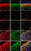
Figure 1.
Figure 1.
Overlapping and differential tissue localization of the N- and C-terminal epitopes of the collagen VI α3 chain. Immunofluorescence microscopy was performed on frozen (a, c, d, and e) or paraffin-embedded sections (b) from murine cornea (a), white adipose tissue (b), muscle (c), skin (d), and knee articular cartilage (e). Shown are adult (a and b) and newborn mice (c–e). Sections were incubated with the affinity-purified antibodies against the collagen VI α3 N terminus (red) and C5 (green); merge is shown in yellow. ep, epithelium, st, stroma. In a, the upper margin of the epithelium is indicated by a dotted line, and in d, the dermal/epidermal basement membrane is shown by a dashed line. Bars, 25 μm in e and 50 μm in a–d.
Figure 2.
Figure 2.
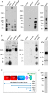
C5-containing fragments of the collagen VI α3 chain are of diverse sizes. Tissue extracts and body fluids were subjected to SDS-PAGE on Tris-glycine 4–12% polyacrylamide gradient gels (a) and on 12% BisTris polyacrylamide gels (b) under nonreducing conditions. Proteins were transferred to a membrane and detected with affinity-purified antibodies against the C5 domain. *, bands that run at the position expected for cleaved off endotrophin. c, domain structure of the C terminus of the collagen VI α3 chain with calculated fragment sizes indicated. FN3, fibronectin-type III repeat; Ku, Kunitz domain. d, mobility of recombinant mouse collagen VI α3 chain C2–C5 fragment (Ala2608–Val3284) on a Tris-glycine 8% polyacrylamide gel. WAT, white adipose tissue.
Figure 3.
Figure 3.
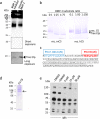
BMP-1 and furin-like proprotein convertase(s) contribute to the processing of the C-terminal part of the collagen VI α3 chain. a, immunoblot of keratocyte conditioned medium incubated with 10 μm BMP-1 inhibitor or 0.27 μg of recombinant BMP-1 (protease/total protein ratio 1:100) for 2 h at 37 °C. Proteins were submitted to SDS-PAGE on a 15% Tris-glycine polyacrylamide gel under nonreducing conditions and detected with an antibody against human endotrophin (C5; Etp). b, cleavage assays with purified recombinant human and mouse collagen VI α3 chain C5 proteins (carrying an N-terminal extension harboring the BMP-1 cleavage site) and BMP-1. Incubation was at 37 °C for 4 h, and detection was by SDS-PAGE on a 4–20% Tris-glycine polyacrylamide gradient gel (reducing conditions) and Coomassie Blue staining. c, summary of the results from the ATOMS experiment performed with the mouse collagen VI α3 C5 protein (rec. mC5). Positions of identified peptides in the protein sequence are shown (P/C, protease/control ratio (mean ± S.D.); the number in brackets indicates the number of peptides used for quantification; underlined is the sequence of the C5 domain as defined in Uniprot; Strep-tag sequence in italics). d, Coomassie Blue–stained SDS-PAGE (8% Tris-glycine polyacrylamide gradient gels under nonreducing conditions) of affinity-purified C-terminally Strep-tagged C1–C5. The arrow indicates the proprotein convertase cleaved band. e, immunoblot of supernatants from primary dermal fibroblasts cultured in the absence or presence of 5, 15, or 30 μm furin inhibitor I for 48 h. The samples were analyzed on a 4–12% Tris-glycine polyacrylamide gradient gel under nonreducing conditions, transferred to a membrane, and detected with the affinity-purified antibody against the mouse collagen VI α3 chain C5 domain.
Figure 4.
Figure 4.
Fibroblasts secrete collagen VI tetramers containing the C5 domain of the collagen VI α3 chain. Primary dermal fibroblasts isolated from newborn mice were cultured for 4 days. a, immunofluorescence microscopy with antibodies against the N terminus (red) and the C5 domain (green) of the collagen VI α3 chain. Bar, 100 μm. b, immunoblot of the collagen VI tetramers in cell lysate (CL) and supernatant (SN) separated under nonreducing conditions on a composite agarose-polyacrylamide (0.5%/2.4%) gel. Purified murine collagen VI tetramers were used as a marker (not shown). The loading buffer contained 2 m urea. Collagen VI tetramers were detected with an affinity-purified antibody against the collagen VI α3 chain C5 domain. t, tetramers.
Figure 5.
Figure 5.
Processing of the C-terminal region of the collagen VI α3 chain occurs after microfibril formation. a, representative collagen VI microfibrils in supernatants of primary fibroblast cultures after 1 and 6 days visualized by EM after negative staining and double labeling using gold-labeled affinity-purified antibodies against the N terminus (10 nm; arrows) and the C5 domain (5 nm; arrowheads) of the collagen VI α3 chain. Bar, 200 nm. Single asterisks indicate the antibody against the C5 domain binding to the triple-helical region; double asterisks indicate antibody binding to C5-containing free particles clearly separated from assembled collagen VI microfibrils. b, statistical evaluation of the location of antibody binding. For each time point, 500 particles from randomly selected areas were counted.

References

    1. Cescon M., Gattazzo F., Chen P., and Bonaldo P. (2015) Collagen VI at a glance. J. Cell Sci. 128, 3525–3531 10.1242/jcs.169748 - DOI - PubMed
    1. Gara S. K., Grumati P., Urciuolo A., Bonaldo P., Kobbe B., Koch M., Paulsson M., and Wagener R. (2008) Three novel collagen VI chains with high homology to the α3 chain. J. Biol. Chem. 283, 10658–10670 10.1074/jbc.M709540200 - DOI - PubMed
    1. Fitzgerald J., Rich C., Zhou F. H., and Hansen U. (2008) Three novel collagen VI chains, α4(VI), α5(VI), and α6(VI). J. Biol. Chem. 283, 20170–20180 10.1074/jbc.M710139200 - DOI - PubMed
    1. Maass T., Bayley C. P., Mörgelin M., Lettmann S., Bonaldo P., Paulsson M., Baldock C., and Wagener R. (2016) Heterogeneity of collagen VI microfibrils: structural analysis of non-collagenous regions. J. Biol. Chem. 291, 5247–5258 10.1074/jbc.M115.705160 - DOI - PMC - PubMed
    1. Gara S. K., Grumati P., Squarzoni S., Sabatelli P., Urciuolo A., Bonaldo P., Paulsson M., and Wagener R. (2011) Differential and restricted expression of novel collagen VI chains in mouse. Matrix Biol. 30, 248–257 10.1016/j.matbio.2011.03.006 - DOI - PubMed

Publication types