. 2019 Jul 26;365(6451):eaav9314.
doi: 10.1126/science.aav9314.

Stem cell differentiation trajectories in Hydra resolved at single-cell resolution

Affiliations

Stem cell differentiation trajectories in Hydra resolved at single-cell resolution

Stefan Siebert et al. Science. .

Abstract

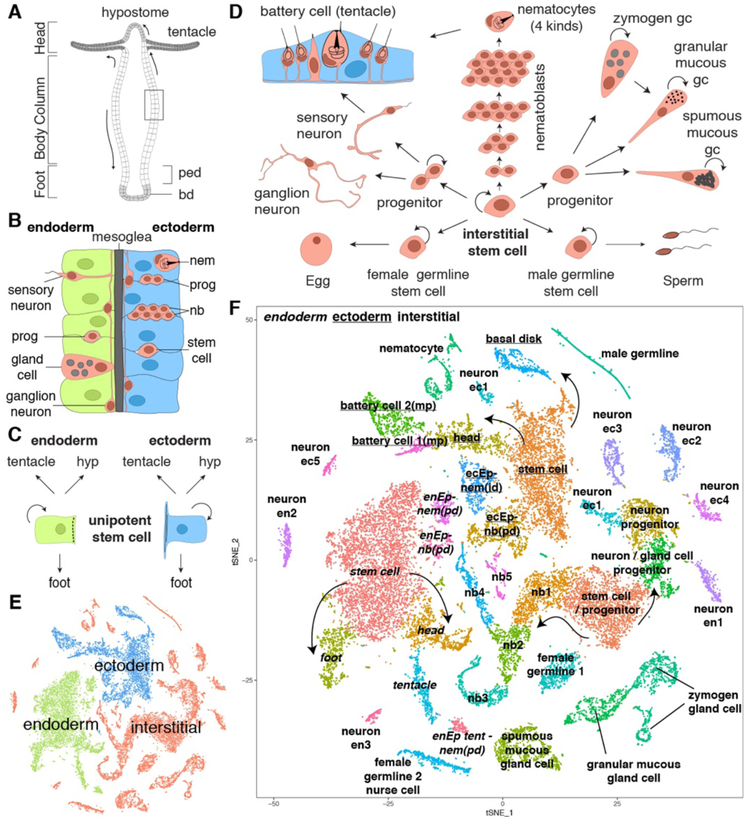
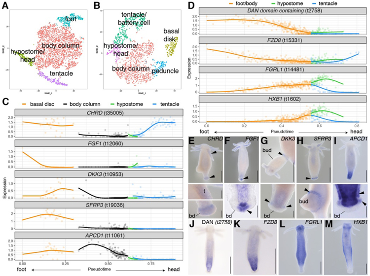
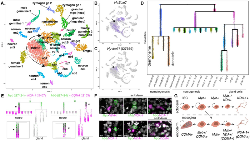
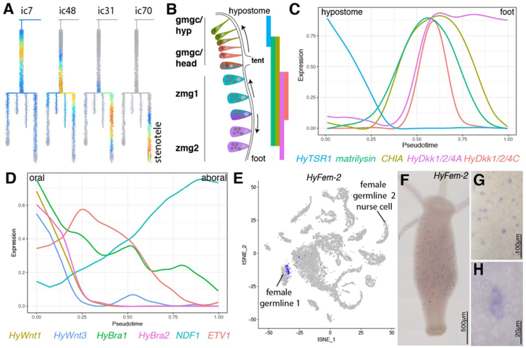
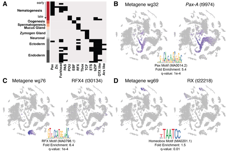
The adult Hydra polyp continually renews all of its cells using three separate stem cell populations, but the genetic pathways enabling this homeostatic tissue maintenance are not well understood. We sequenced 24,985 Hydra single-cell transcriptomes and identified the molecular signatures of a broad spectrum of cell states, from stem cells to terminally differentiated cells. We constructed differentiation trajectories for each cell lineage and identified gene modules and putative regulators expressed along these trajectories, thus creating a comprehensive molecular map of all developmental lineages in the adult animal. In addition, we built a gene expression map of the Hydra nervous system. Our work constitutes a resource for addressing questions regarding the evolution of metazoan developmental processes and nervous system function.


Conflict of interest statement

Competing interests: The authors declare no competing interests.

Figures

Figure 1.
Figure 1.. Hydra tissue composition and single cell RNA sequencing of 24,985 Hydra cells.
A) The Hydra body is a hollow tube with an adhesive foot at the aboral end (bd: basal disk, ped: peduncle) and a head with a mouth and a ring of tentacles at the oral end. The mouth opening is at the tip of a cone shaped protrusion — the hypostome. B) Enlargement of box in A. The body column consists of two epithelial layers (endoderm and ectoderm) separated by an extracellular matrix — the mesoglea. Cells of the interstitial cell lineage (red) reside in the interstitial spaces between epithelial cells, except gland cells which are integrated into the endodermal epithelium. Ectodermal cells can enclose nerve cells or nematocytes forming biological doublets. C) Epithelial cells of the body column are mitotic, have stem cell properties, and give rise to terminally differentiated cells of the hypostome (hyp), tentacles, and foot. D) Schematic of the interstitial stem cell lineage. The lineage is supported by a multipotent interstitial stem cell (ISC) that gives rise to neurons, gland cells, and nematocytes; ISCs are also capable of replenishing germline stem cells if they are lost. E) t-SNE representation of clustered cells colored by cell lineage. F) t-SNE representation of clustered cells annotated with cell state. Figures A-D adapted from (50). ec: ectodermal, en: endodermal, Ep: epithelial cell, gc: gland cell, id: integration doublet, mp: multiplet, nb: nematoblast, nem: differentiated nematocyte, pd: suspected phagocytosis doublet. id, mp, and pd are categories of biological doublets. Arrows indicate suggested transitions from stem cell populations to differentiated cells.
Figure 2.
Figure 2.. Identification of genes with differential expression along the oral-aboral axis.
A) t-SNE representation of subclustered endodermal epithelial cells and B) subclustered ectodermal epithelial cells. C-D) Epithelial cells were ordered using URD to reconstruct a trajectory where pseudotime represents spatial position. Scaled and log-transformed expression is visualized. C) Trajectory plots for previously uncharacterized putative signaling genes expressed in ectodermal epithelial cells of foot and tentacles. BMP antagonist CHRD (t35005), fibroblast growth factor FGF1 (t12060); Wnt antagonists DKK3 (t10953), SFRP3 (t19036) and APCD1 (t11061). D) Trajectory plots for genes expressed in a graded manner in endodermal epithelial cells. BMP antagonist “DAN domain containing gene” t2758, secreted Wnt antagonist FZD8 (t15331), fibroblast growth factor receptor FGRL1 (t14481), homeobox protein HXB1 (t1602). E-M) Epithelial expression patterns obtained using RNA in situ hybridization consistent with predicted patterns. Whole mounts and selected close-ups. Arrows indicate ectodermal signal. t: tentacle, bd: basal disk. Scale bars: whole mounts (including G): 500 μm, close-ups: 100 μm.
Figure 3.
Figure 3.. Trajectory reconstruction for cells of the interstitial lineage suggests a cell state common to neurogenesis and gland cell differentiation.
A) t-SNE representation of interstitial cells with clusters labeled by cell state. Solid arrow: neurogenesis/gland cell differentiation. Dashed arrow: nematogenesis. B) HvSoxC expression in progenitor cells. Arrow indicates putative ISC population, which is negative for HvSoxC and positive for biomarker Hy-icell1 expression (C). D) URD differentiation tree of the interstitial lineage. Color represents URD segments and do not correspond to the colors in the t-SNE (see fig. S19. E)) Myb (green) is expressed in the neuron/gland cell progenitor state and during early neurogenesis/gland cell differentiation. Expression of Myb (green, > 0) partially overlaps with high expression of the neuronal gene NDA-1 (magenta, > 3) and the gland cell gene COMA (t2163) (magenta, > 0); COMA is also expressed in a subset of endodermal neurons. Co-expressing cells are black. Star and close-up highlights cell states with co-expression. F) Double labeling using fluorescent RNA in situ hybridization is consistent with neuron differentiation in the endodermal and ectodermal epithelial layers and demonstrates the existence of transition states observed in the trajectory analysis. Additionally, endodermal gland cell differentiation transition states were observed in the endodermal epithelial layer (see also fig. S22). gc: gland cell, gp: gland cell progenitor, n: neuron, np: neuron progenitor, p: Myb positive progenitor. G) Model for progenitor specification. Ectodermal ISCs give rise to a progenitor that can give rise to ectodermal neurons. Progenitors that translocate to the endoderm are able to give rise to glands or neurons. ec: ectoderm, en: endoderm, gmgc: granular mucous gland cell, gc: gland cell, hyp: hypostome, ISC: interstitial multipotent stem cell, mgc: mucous gland cell, nb: nematoblast, smgc: spumous mucous gland cell, prog: progenitor, zmg: zymogen gland cell.
Figure 4.
Figure 4.. Subtrajectory analyses of interstitial cell types.
A) Interstitial gene modules successively expressed in nematocytes forming a stenotele (ic: interstitial gene module). B) Model for gland cell (ZMG/gMGC) location dependent changes. Gland cells integrated in the endodermal epithelium get displaced towards the extremities and undergo changes in expression and morphology. Bars show known expression domains for genes depicted in (C). gmgc: granular mucous gland cell, hyp: hypostome, tent: tentacle, zmg: zymogen gland cell. C) URD linear ZMG/gMGC trajectory recapitulates known position dependent gene expression in gland cells along the body column. HyTSR1 (15), HyDkk1/2/4 A/C (51, 52), matrilysin-like (t32151)/CHIA (t18356) (fig. S26B–E). D) URD linear sMGC trajectory plot for HyWnt1 (53), HyWnt3 (54), Hybra1 (55), Hybra2 (56), ETV1 (t22116) and NDF1 (t21810) showing expression changes in pseudotime that correlate to position along the oral-aboral axis. Cells are ordered according to pseudotime with putative hypostomal cell states to the left and putative lower head cell states to the right. E) Plot showing HyFem-2 expression in a subset of cells in the early female cluster. F-H) HyFem-2 is expressed in single cells or pairs scattered within the body column.
Figure 5.
Figure 5.. Motif enrichment analysis for gene modules and identification of candidate regulators.
A) Enriched motifs (columns) found in open chromatin of putative 5’ cis-regulatory regions of co-expressed gene sets (metagenes) for listed cell states (rows). B-D) Metagene scores visualized on the t-SNE representation (left), significantly enriched motif found in putative 5’ cis-regulatory regions (bottom) and candidate regulators likely to bind identified motif with correlated expression (right). B) Metagene expressed during nematogenesis and putative PAX regulator. C) Metagene expressed in gland cells and putative RFX regulator. D) Metagene expressed in ectodermal epithelial cells of the foot and putative homeobox regulator.
Figure 6.
Figure 6.. Molecular map of the Hydra nervous system with spatial resolution.
A) Subclustering of neurons and neuronal progenitors. Cell states are annotated with cell layer, localization along the body column, tentative neuronal subtype category — sensory (S) or ganglion (G), and gene markers used in annotations. B) Heatmap shows top twelve markers for neuronal cell states. C-E) First molecular markers for endodermal neurons. C) Transgenic line (NDF1(t14976)::GFP) expressing GFP in endodermal ganglion neurons along the body column (cluster “en1”). D-E) Body column cross-section of transgenic line (Alpha-LTX-Lhe1a-like(t33301)::GFP) expressing GFP in putative sensory neurons (cluster “en2”). Phalloidin staining (red) marks ECM and Hoechst (blue) marks nuclei. en: endoderm, ec: ectoderm.

Comment in

  • The cells of regeneration.
    Reddien PW. Reddien PW. Science. 2019 Jul 26;365(6451):314-316. doi: 10.1126/science.aay3660. Science. 2019. PMID: 31346049 No abstract available.
  • Looking at Hydra cells one at a time.
    Vogt N. Vogt N. Nat Methods. 2019 Sep;16(9):801. doi: 10.1038/s41592-019-0569-6. Nat Methods. 2019. PMID: 31471615 No abstract available.
  • Many Ways to Build a Polyp.
    Arendt D. Arendt D. Trends Genet. 2019 Dec;35(12):885-887. doi: 10.1016/j.tig.2019.09.003. Epub 2019 Oct 16. Trends Genet. 2019. PMID: 31629552

References

    1. Trembley A, Mémoires Pour Servirà l’Histoire d’un Genre de Polypes d’Eau Douce, à Bras en Forme de Cornes (Chez Jean & Herman Verbeek, Leiden, 1744).
    1. Weismann A, Die Entstehung der Sexualzellen bei den Hydromedusen: Zugleich ein Beitrag zur Kenntniss des Baues und der Lebenserscheinungen dieser Gruppe (Gustav Fischer Verlag, Jena, 1883).
    1. Campbell RD, Development of Hydra Lacking Interstitial and Nerve Cells (“Epithelial Hydra”) in Determinants of Spatial Organization (Elsevier, 1979), pp. 267–293.
    1. Sugiyama T, Fujisawa T, Genetic analysis of developmental mechanisms in Hydra. II. Isolation and characterization of an interstitial cell-deficient strain. J. Cell. Sci 29, 35–52 (1978). - PubMed
    1. David CN, Murphy S, Characterization of interstitial stem cells in Hydra by cloning. Dev. Biol 58, 372–383 (1977). - PubMed

Publication types