. 2019 Jul 25;9(1):10782.
doi: 10.1038/s41598-019-47209-1.

Phylogeographic and genetic characterization of porcine circovirus type 2 in Taiwan from 2001-2017

Affiliations

Phylogeographic and genetic characterization of porcine circovirus type 2 in Taiwan from 2001-2017

Guang-Ting Tsai et al. Sci Rep. .

Abstract

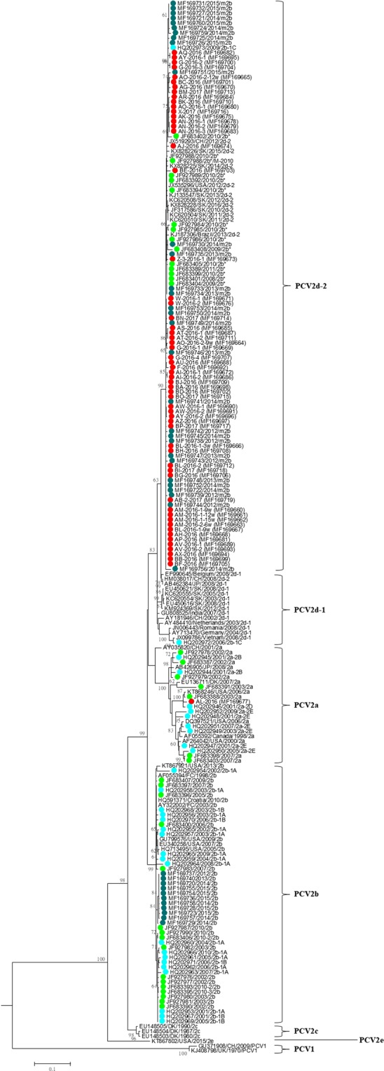
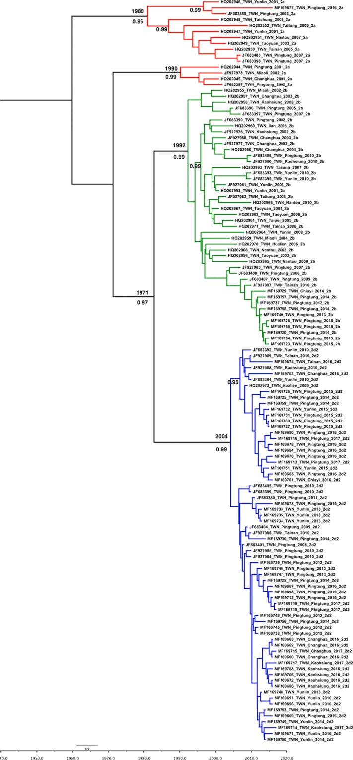
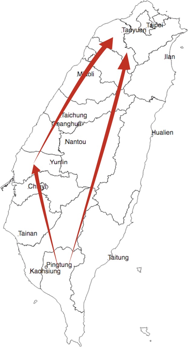
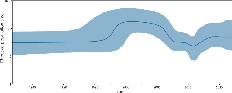
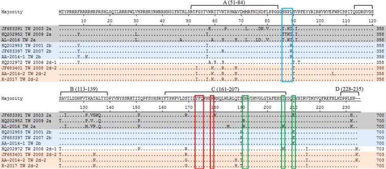
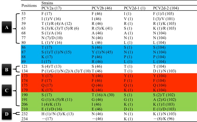
Porcine circovirus type 2 (PCV2) is an important pathogen that causes significant economic losses in the swine industry worldwide. Five major PCV2 genotypes have been identified, including PCV2a, PCV2b, PCV2c, PCV2d, and PCV2e. To investigate the prevalence and phylodynamics of the different PCV2 genotypes in Taiwan, 214 PCV2 ORF2 sequences from Taiwan and other countries were analyzed. Genotypic differences were observed among PCV2a, 2b, and 2d at amino acid position 89 in ORF2, with isoleucine (I), arginine (R), and leucine (L), respectively. Similar to other countries, a genotypic shift was also observed in Taiwan, where the predominant genotype shifted from PCV2b to 2d after 2010. The estimated nucleotide substitution rate of Taiwanese strains in the ORF2 region was 8.467 × 10⁻⁴ substitutions per site per year. This rapid evolution rate of PCV2 may lead to the genotypic shift observed in Taiwan. The times to the most recent common ancestor (TMRCA) for PCV2a, -2b, and -2d-2 was dated to 1970, 1992 and 2004, respectively. Thus, the PCV2a, -2b, and -2d genotypes were already present in Taiwan before the introduction of the PCV2 vaccine.


Conflict of interest statement

The authors declare no competing interests.

Figures

Figure 1
Figure 1
PCV2 positivity rate in swine samples deposited at the ADDC of NPUST in Taiwan from July 2014 to December 2016. The numbers above bars indicate the number of PCV2-positive pigs out of the total number of pigs in which detection was attempted.
Figure 2
Figure 2
Detection of histopathological lesions in typical PCVAD clinical cases by hematoxylin-eosin staining. (A,B) Necrotizing lesion of the submandibular lymph node with basophilic endoplasmic inclusion bodies in mononuclear cells. (C) The alveolar septa were thickened by interstitial infiltration of mononuclear inflammatory cells. (D) The mesenteric lymph node contained basophilic endoplasmic inclusion bodies in mononuclear cells.
Figure 3
Figure 3
PCV2 immunochemistry of typical PCVAD clinical cases. (A) The interstitial pneumonia lesion contained PCV2 antigen positive signals (brown) within the cytoplasm of macrophage-like cells. (B) PCV2 immunolabeling (brownish signal) is seen in the cytoplasm of macrophage-like cells in the lymph node.
Figure 4
Figure 4
Phylogenetic tree based on PCV2 ORF2 gene sequences. The tree was constructed using the maximum-likelihood method based on the general time reversible (GTR) model with a gamma-distributed rate (five rate categories) and an invariant site (I). Taiwanese PCV2 strains were labeled with solid dots of different colors: red, blue-green, bright-green, and bright-blue dots represent samples collected from 2016–2017, 2012–2015, 2002–2011, and 2001–2010, respectively.
Figure 5
Figure 5
Maximum-clade-credibility (MCC) tree of PCV2 inferred from 126 complete ORF2 sequences from Taiwan. The MCC tree was constructed with a 10% burn-in using Tree Annotator v 1.10 implemented in the BEAST software package. The red, green and blue branch lines represent the PCV2a, PCV2b, and PCV2d isolates, respectively. The numbers adjacent to the branches are the posterior probability values and branch times. Only posterior probability values greater than 0.95 are shown.
Figure 6
Figure 6
Migration of temporal dynamics for PCV2 in Taiwan. The visualizing location annotated Maximum Clade Credibility tree by SPREAD3 software. The branches show an overview of the possible transmission routes of PCV2 in Taiwan. The direction of the spread of the virus is indicated with arrows.
Figure 7
Figure 7
Bayesian skyline plot for the complete capsid gene encoding PCV2. The x-axis shows the years before 2017, while the y-axis represents effective viral population size. The thicker bold line represents the median estimate of the effective number of infections over time, whereas the thinner blue lines indicate the upper and lower bounds of the 95% HPD.
Figure 8
Figure 8
Amino acids of the putative PCV2 capsid protein for PCV2a (gray), PCV2b (blue), and PCV2d-1 and PCV2d-2 (orange). The four general antibody recognition regions are labeled (A) (51–84), (B) (113–139), (C) (161–207), and (D) (228–233). Positions 86–89 (blue box) were previously proposed to distinguish between PCV2a and PCV2b. Positions 190–191, 206, and 210 (green box) are important for PCV2 replication in vitro. Positions 173–175 and 179 (red box) are important for antibody recognition.
Figure 9
Figure 9
Variable residues in the putative capsid protein of ORF2 among 168 PCV2 isolates from Taiwan. Amino acid respective number was represented for amount of strains. Four general antibody recognition regions were labeled (A) (51–84), (B) (113–139), (C) (161–207), and (D) (228–233). Positions 86–89 (blue) were proposed to distinguish between PCV2a and PCV2b. Positions 173–175 and 179 (red) are important for antibody recognition. Positions 190–191, 206, and 210 (green) are important for PCV2 replication in vitro.

References

    1. Opriessnig T, Meng XJ, Halbur PG. Porcine circovirus type 2 associated disease: Update on current terminology, clinical manifestations, pathogenesis, diagnosis, and intervention strategies. J. Vet. Diagn. Invest. 2007;19:591–615. doi: 10.1177/104063870701900601. - DOI - PubMed
    1. Segalés J. Porcine circovirus type 2 (PCV2) infections: clinical signs, pathology and laboratory diagnosis. Virus Res. 2012;164:10–19. doi: 10.1016/j.virusres.2011.10.007. - DOI - PubMed
    1. Segalés J, Allan GM, Domingo M. Porcine circovirus diseases. Anim. Health Res. Rev. 2005;6:119–142. doi: 10.1079/AHR2005106. - DOI - PubMed
    1. Cheung AK. The essential and nonessential transcription units for viral protein synthesis and DNA replication of porcine circovirus type 2. Virology. 2003;313:452–459. doi: 10.1016/S0042-6822(03)00373-8. - DOI - PubMed
    1. Mankertz J, Buhk HJ, Blaess G, Mankertz A. Transcription analysis of porcine circovirus (PCV) Virus Genes. 1998;16:267–276. doi: 10.1023/A:1008022521329. - DOI - PubMed

LinkOut - more resources