. 2019 Nov;224(8):2661-2676.
doi: 10.1007/s00429-019-01922-9. Epub 2019 Jul 25.

Age-related hearing loss increases full-brain connectivity while reversing directed signaling within the dorsal-ventral pathway for speech

Affiliations

Age-related hearing loss increases full-brain connectivity while reversing directed signaling within the dorsal-ventral pathway for speech

Gavin M Bidelman et al. Brain Struct Funct. 2019 Nov.

Abstract

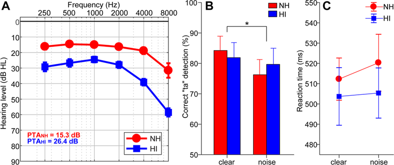
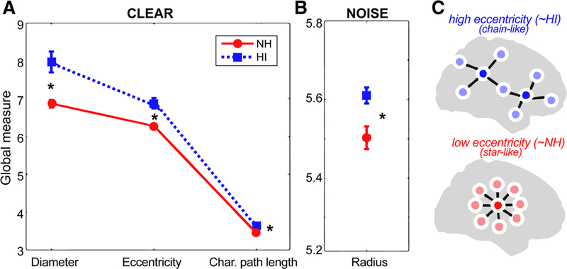
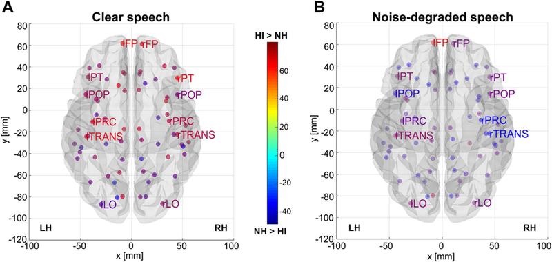
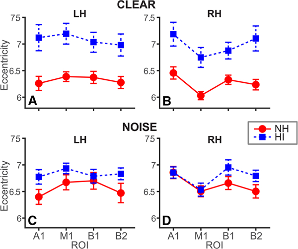
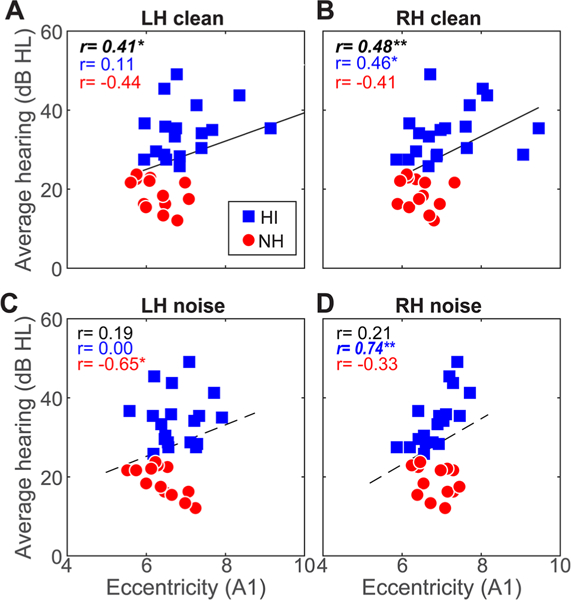
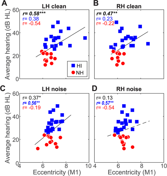
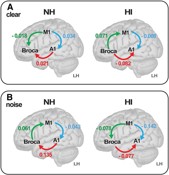
Speech comprehension difficulties are ubiquitous to aging and hearing loss, particularly in noisy environments. Older adults' poorer speech-in-noise (SIN) comprehension has been related to abnormal neural representations within various nodes (regions) of the speech network, but how senescent changes in hearing alter the transmission of brain signals remains unspecified. We measured electroencephalograms in older adults with and without mild hearing loss during a SIN identification task. Using functional connectivity and graph-theoretic analyses, we show that hearing-impaired (HI) listeners have more extended (less integrated) communication pathways and less efficient information exchange among widespread brain regions (larger network eccentricity) than their normal-hearing (NH) peers. Parameter optimized support vector machine classifiers applied to EEG connectivity data showed hearing status could be decoded (> 85% accuracy) solely using network-level descriptions of brain activity, but classification was particularly robust using left hemisphere connections. Notably, we found a reversal in directed neural signaling in left hemisphere dependent on hearing status among specific connections within the dorsal-ventral speech pathways. NH listeners showed an overall net "bottom-up" signaling directed from auditory cortex (A1) to inferior frontal gyrus (IFG; Broca's area), whereas the HI group showed the reverse signal (i.e., "top-down" Broca's → A1). A similar flow reversal was noted between left IFG and motor cortex. Our full-brain connectivity results demonstrate that even mild forms of hearing loss alter how the brain routes information within the auditory-linguistic-motor loop.

Keywords: EEG; Functional connectivity; Global and nodal network features; Graph theory; Hearing loss; Machine learning.


Conflict of interest statement

Conflict of interest The authors declare they have no conflict of interest.

Figures

Fig. 1
Fig. 1
Behavioral data. a Audiograms of normal-hearing (NH) and hearing-impaired (HI) listeners. b Behavioral accuracy for detecting infrequent /ta/ tokens in clear and noise-degraded conditions. Noise-related decline in behavioral performance was prominent but no group differences in speech perception were observed. c Reaction times (RTs) for speech detection were similar between groups and speech SNRs. Error bars = ± s.e.m., *p < 0.05
Fig. 2
Fig. 2
Group comparison of mean global brain connectivity measures during clear and noise-degraded speech processing. a Graph measures of diameter, eccentricity, and characteristic path length were larger in HI compared to NH listeners when processing clear speech. b For noise-degraded speech, only radius measures of the network differed between groups. c Schematic of two functional brain networks varying in eccentricity (see Fig. 3 for raw data). High eccentricity networks (like that of HI listeners) have more chain-like global configuration reflecting less integration and more long-range neural signaling; low eccentricity networks (like that of NH listeners) have configurations that are more integrated and “star-like.” After He et al. (2018). See text for definitions of graph metrics. *p < 0.05, error bars = ± s.e.m
Fig. 3
Fig. 3
Group contrast of full-brain nodal eccentricity as a function of speech SNR. a Clear speech. b Noise-degraded speech. Hotter colors denote nodes of the DK atlas (Desikan et al. 2006) where HI listeners showed larger interconnectivity (eccentricity) between network nodes [(i.e., HI > NH); cooler colors, NH > HI]. Note the stronger eccentricity in the HI group in frontal and temporal sites. Functional data are projected onto the BRAPH glass brain template (Mijalkov et al. 2017). For clarity, only select DK ROIs are labeled. FP, frontal pole; LO, lateral occipital; POP, pars opercularis (i.e., “B1”); PT, pars triangularis (i.e., “B2”); PC, precentral gyrus (i.e., “M1”); TRANS, transverse temporal gyrus (i.e., “A1”)
Fig. 4
Fig. 4
Age-related hearing loss is associated with more extended pathways of neural communication, particularly in the left hemi-sphere. Average eccentricity within specific nodes of the auditory-linguistic-motor loop in left (LH) and right (RH) hemispheres. a, b Clear speech. c, d Noise-degraded speech. While HI listeners show increased network eccentricity, group differences are largest in LH. A1, primary auditory cortex; M1, primary motor cortex; B1-B2, Broca’s area (pars opercularis and pars triangularis). Error bars = ± s.e.m
Fig. 5
Fig. 5
Connectivity in/outflow of auditory cortex is associated with hearing acuity. a, b Correlations between A1 connectivity during clear speech processing and hearing thresholds. Both LH and RH connectivity covaries with hearing acuity; larger eccentricity is linked with more severe hearing loss. c, d A1 eccentricity in the noise condition is not related to audiometric thresholds when considering all listeners. Solid lines, significant correlations; dotted lines, n.s. Bold italics = correlations surviving FDR correction across SNR and hemisphere. *p < 0.05, **p < 0.01, ***p < 0.001
Fig. 6
Fig. 6
Connectivity in/outflow of motor cortex is associated with hearing acuity for both clear and noise-degraded speech. Hearing loss is predicted by M1 connectivity in both LH and RH for clear and noise-degraded speech. Otherwise as in Fig. 5
Fig. 7
Fig. 7
Directional flow of neural signaling within the dorsal–ventral stream reverses with age-related hearing loss. a Clear speech. b Noise-degraded speech. Values represent the strength of connectivity within LH computed via phase transfer entropy (Lobier et al. 2014), whereas the direction (causality) of communication is determined by sign. Arrows denote flow from region A → B. The NH listeners show signaling directed from A1 → Broca’s (pars opercularis), whereas the HI group shows the reverse (Broca’s → A1), suggesting bottom-up versus top-down configurations within the same pathway dependent on hearing status. During noise-degraded speech, communication between linguistic and motor areas reverses from Broca’s driving M1 to M1 driving Broca’s, but only in HI listeners (cf. green connection, A vs. B)

References

    1. Abdala C, Dhar S (2012) Maturation and aging of the human cochlea: a view through the DPOAE looking glass. J Assoc Res Otolaryngol 13(3):403–421 - PMC - PubMed
    1. Alain C, Snyder JS (2008) Age-related differences in auditory evoked responses during rapid perceptual learning. Clin Neurophysiol 119(2):356–366 - PubMed
    1. Alain C, McDonald K, Van Roon P (2012) Effects of age and back-ground noise on processing a mistuned harmonic in an otherwise periodic complex sound. Hear Res 283:126–135 - PubMed
    1. Alain C, Roye A, Salloum C (2014) Effects of age-related hearing loss and background noise on neuromagnetic activity from auditory cortex. Front Syst Neurosci 8:8. - PMC - PubMed
    1. Alain C, Arsenault JS, Garami L, Bidelman GM, Snyder JS (2017) Neural correlates of speech segregation based on formant frequencies of adjacent vowels. Sci Rep 7(40790):1–11 - PMC - PubMed