. 2019 Jul 25;14(1):250.
doi: 10.1186/s11671-019-3080-1.

Oxidizing and Nano-dispersing the Natural Silk Fibers

Affiliations

Oxidizing and Nano-dispersing the Natural Silk Fibers

Ke Zheng et al. Nanoscale Res Lett. .

Abstract

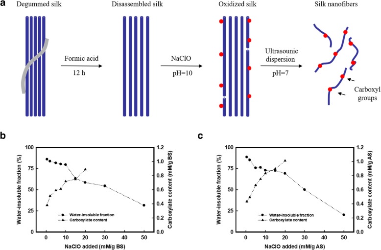
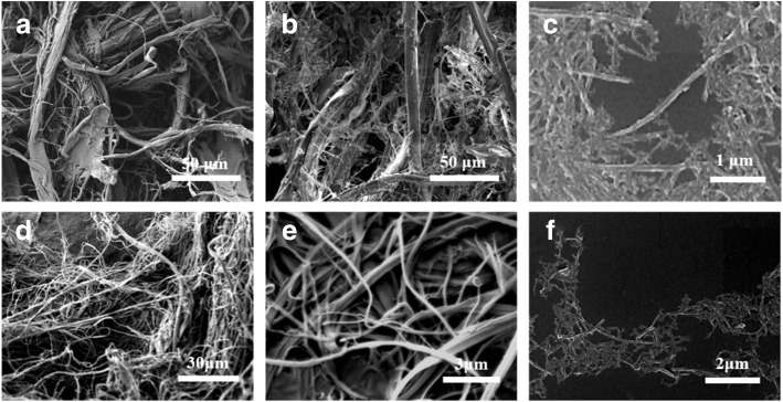
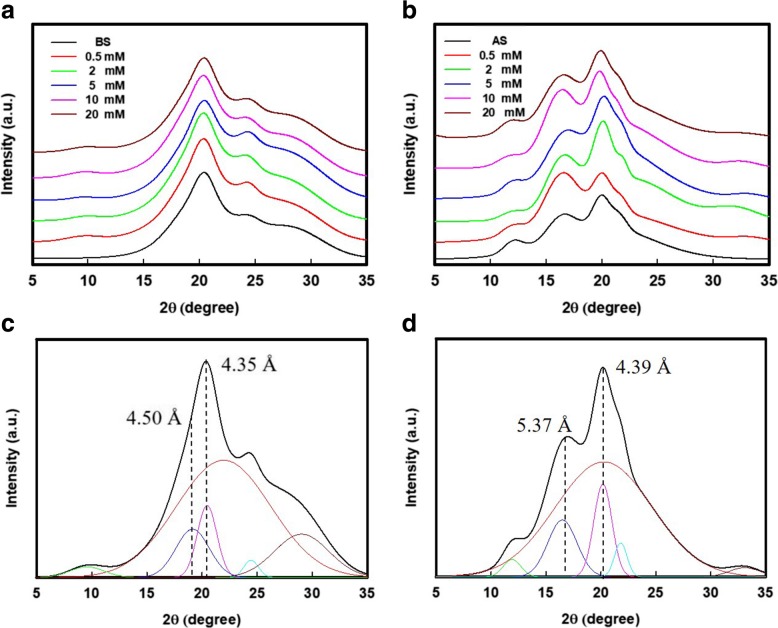
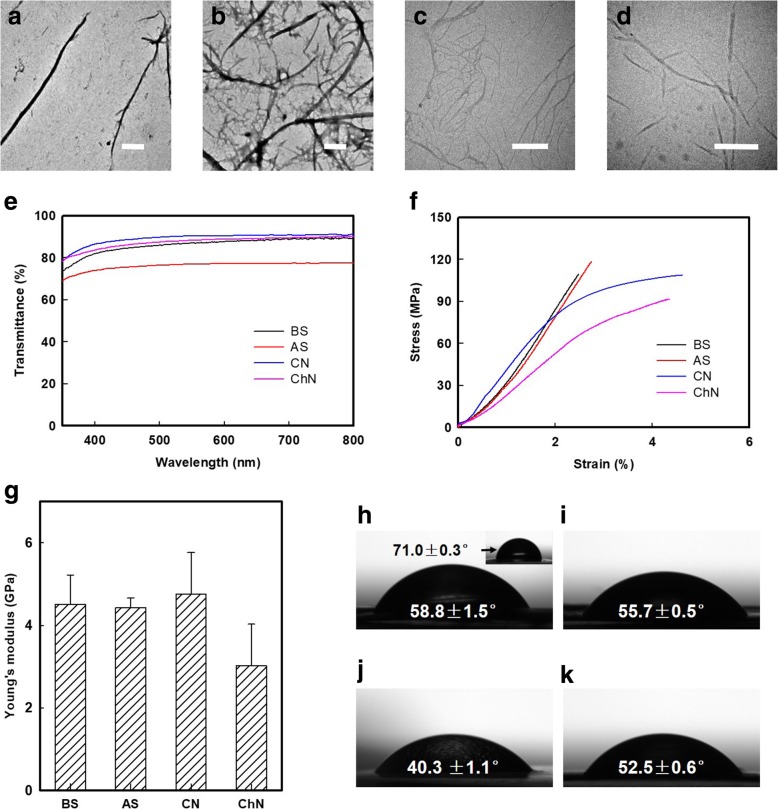
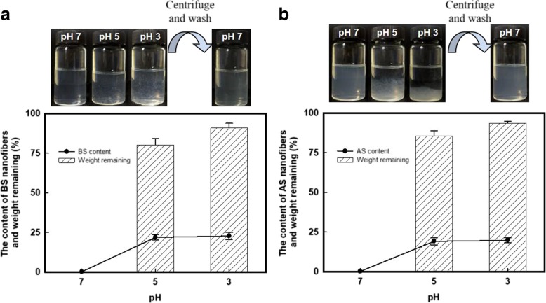
Natural Bombyx mori silk (BS) and Antheraea pernyi silk (AS) were oxidized in sodium hypochlorite (NaClO) solutions. Thereafter, individual silk nanofibers (SNs) were achieved after sonicating the oxidized silk slurries, where the diameters of the resultant SNs were ~ 100 nm and several micrometers in length. Thin membranes were formed by casting the SNs, which had optically transparent (above 75% transmission), mechanically robust (~4.5 GPa of Young's modulus), and enhanced wetting properties. An interesting aggregating-dispersing (re-dispersing) process by using these SNs was strongly regulated by adjusting the pH values. Consequently, the negatively charged SNs could be concentrated up to ~ 20 wt% (100 times that of the initial dispersion) and offered extraordinary benefits for storage, transportation, and engineering applications.

Keywords: Aggregating-redispersing; Negatively charged nanofibers; Oxidation; Silk.


Conflict of interest statement

The authors declare that they have no competing interests.

Figures

Fig. 1
Fig. 1
Process diagram of SNs and carboxyl content of BS and AS. a Schematic of oxidation and dispersing of silk fiber to silk nanofibers (SNs). b The content of carboxyl groups and weight remaining of water-insoluble fraction after oxidation of Bombyx mori (BS) corresponding to the addition of sodium hypochlorite (NaClO). The carboxyl content increased from 0.293 to 0.889 mM/g BS (NaClO addition was 20 mM/g protein) with 58.52 wt% of protein remaining. c For Antheraea pernyi silk (AS). The carboxyl content increased from 0.347 to 1.013 mM/g AS (NaClO addition was 20 mM/g protein) with 69.30 wt% of protein remaining
Fig. 2
Fig. 2
Representative SEM observation of resultant silk fibers in each process. a Disassembled BS fibers after formic acid pretreatment, b oxidized BS fibers, and c the BS nanofibers with a diameter of 105 ± 27 nm. d Disassembled AS fibers after formic acid pretreatment, e oxidized AS fibers, and f the AS nanofibers with a diameter of 112 ± 33 nm. The contour length of BS and AS nanofibers is more than 1 μm
Fig. 3
Fig. 3
XRD analysis of the oxidized silk fibers. X-ray diffraction (XRD) pattern of a BS and b AS that oxidized with various NaClO addition. Representative deconvolution and results of c BS and d AS materials
Fig. 4
Fig. 4
Morphology and properties testing of SNs, CNs, and ChNs. Transmission electron microscopy (TEM) observation of resultant a BS and b AS nanofibers that oxidized by 10 mM/g protein NaClO addition, c cellulose nanofibers (CNs), and d chitin nanofibers (ChNs) that achieved by TEMPO-mediated oxidation. The scale bar is 500 nm. e UV-Vis transmittance of approximately 50-μm-thick membranes that cast by BS, AS, cellulose (CN), and chitin (ChN) nanofibers. f Representative stress-strain curves of approximately 50-μm-thick membranes that cast by BS, AS, CN, and ChN nanofibers. g Young’s modulus of membranes that are casting from BS, AS, CN, and ChN nanofibers. Data represent the mean SD (n = 5). hk The water contact angle of membrane cast by f BS nanofibers was 58.8 ± 1.5°, significantly reduced from that of regenerated BS membrane (71.0 ± 0.3°, the inset image). 55.7 ± 0.5, 40.3 ± 1.1, and 52.5 ± 0.6° of water contact angle was presented in AS, CN, and ChN membrane, respectively
Fig. 5
Fig. 5
Re-dispersing process of SNs. Photography of the pH response phenomenon for a BS and b AS nanofibers. Over 80 wt% of the proteins (both BS and AS) were remaining after centrifugation, with the protein content of ~ 20 wt%

References

    1. Fratzl P, Weinkamer R. Nature’s hierarchical materials. Prog Mater Sci. 2007;52:1263–1334. doi: 10.1016/j.pmatsci.2007.06.001. - DOI
    1. Mano J, Silva G, Azevedo H, et al. Natural origin biodegradable systems in tissue engineering and regenerative medicine: present status and some moving trends. J R Soc Interface. 2007;4:999–1030. doi: 10.1098/rsif.2007.0220. - DOI - PMC - PubMed
    1. Lakes R. Materials with structural hierarchy. Nature. 1993;361:511–515. doi: 10.1038/361511a0. - DOI
    1. Wegst UGK, Bai H, Saiz E, et al. Bioinspired structural materials. Nat Mater. 2015;14:23–36. doi: 10.1038/nmat4089. - DOI - PubMed
    1. Mishnaevsky L, Tsapatsis M. Hierarchical materials: background and perspectives. MRS Bull. 2016;41:661–664. doi: 10.1557/mrs.2016.189. - DOI