Review
. 2019 Jul;79(7):639-655.
doi: 10.1002/dneu.22712. Epub 2019 Aug 6.

Exosome release and cargo in Down syndrome

Affiliations
Review

Exosome release and cargo in Down syndrome

Eric D Hamlett et al. Dev Neurobiol. 2019 Jul.

Abstract

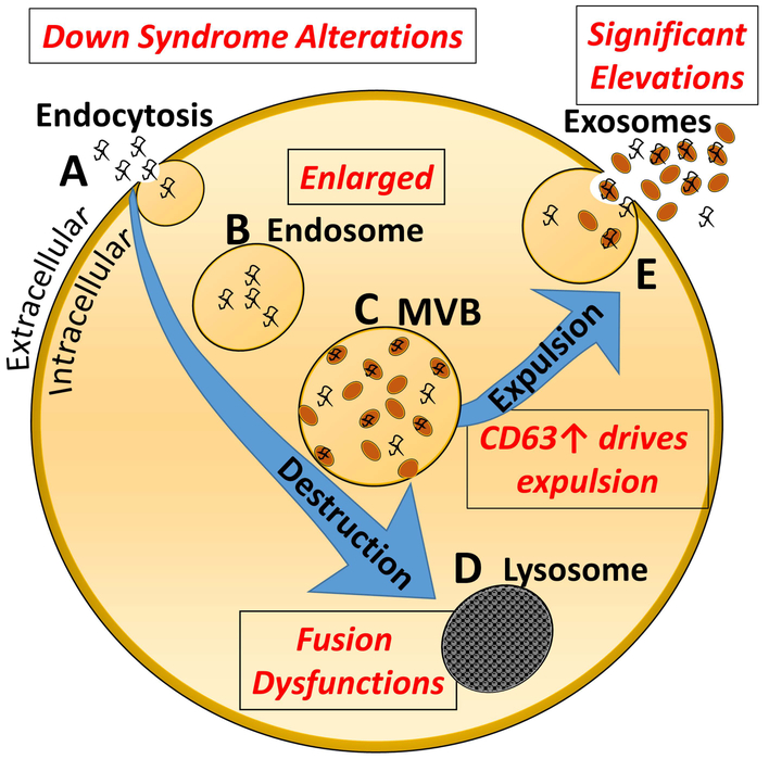
Down syndrome (DS) is a multisystem disorder affecting 1 in 800 births worldwide. Advancing technology, medical treatment, and social intervention have dramatically increased life expectancy, yet there are many etiologies of this disorder that are in need of further research. The advent of the ability to capture extracellular vesicles (EVs) in blood from specific cell types allows for the investigation of novel intracellular processes. Exosomes are one type of EVs that have demonstrated great potential in uncovering new biomarkers of neurodegeneration and disease, and also that appear to be intricately involved in the transsynaptic spread of pathogenic factors underlying Alzheimer's disease and other neurological diseases. Exosomes are nanosized vesicles, generated in endosomal multivesicular bodies (MVBs) and secreted by most cells in the body. Since exosomes are important mediators of intercellular communication and genetic exchange, they have emerged as a major research focus and have revealed novel biological sequelae involved in conditions afflicting the DS population. This review summarizes current knowledge on exosome biology in individuals with DS, both early in life and in aging individuals. Collectively these studies have demonstrated that complex multicellular processes underlying DS etiologies may include abnormal formation and secretion of extracellular vesicles such as exosomes.

Keywords: Alzheimer's disease; Down syndrome; biomarkers; extracellular vesicles; neurodegeneration.


Conflict of interest statement

Declaration of Interest: The authors have no conflict of interest with companies or other organizations that can bias this review.

Figures

Figure 1.
Figure 1.. Exosome biogenesis and secretion.
A. Invagination of the plasma membrane into signaling endosomes. B. Transport of the vesicle into a larger endosome. C. The endosome will mature into a multivesicular body (MVB). D. The MVB undergoes inward budding, generating, and accumulating intraluminal vesicles that can either be directed to the lysosome for degradation (E) or fused to the plasma membrane for subsequent release (F). Alterations noted in the DS population, or in DS mouse models, are shown in Red in the figure.
Figure 2.
Figure 2.. Exosome purification method.
A. Plasma or serum samples are obtained and the Exoquick polymer employed to obtain total exosomes from the fluid. Thereafter, an L1CAM antibody capture system is utilized to purify all exomes of neuronal origin from the sample. B. CD81 levels in DS and control NDEs. Multiple blood samples at each age from DS and a typically developing population were obtained, exosomes purified and CD81 levels measured by ELISA. As can be seen here, most DS samples contained elevated amounts of CD81, indicating an increase in exosome release in this population at all ages analyzed. [Original data Hamlett, Ledreux and Granholm et al]
Figure 3.
Figure 3.. L1CAM immunostaining in various organs in human post mortem tissues.
L1CAM immunostaining is enriched in (A) cortical tissues, and is much less abundant to near absent in (B-F) other organs. Thus, this antibody represents an excellent tool for immunoprecipitation of brain-derived exosomes, as used in our protocols. Photos were obtained from the Human Protein Atlas which is licensed under the Creative Commons Attribution-ShareAlike 3.0 International License (thus not copyrighted). From Uhlen et al., 2015.

References

    1. Abdulrahman BA, Abdelaziz DH, Schatzl HM, 2018. Autophagy regulates exosomal release of prions in neuronal cells. J Biol Chem 293, 8956–8968. - PMC - PubMed
    1. Annus T, Wilson LR, Hong YT, Acosta-Cabronero J, Fryer TD, Cardenas-Blanco A, Smith R, Boros I, Coles JP, Aigbirhio FI, Menon DK, Zaman SH, Nestor PJ, Holland AJ, 2016. The pattern of amyloid accumulation in the brains of adults with Down syndrome. Alzheimers Dement 12, 538–545. - PMC - PubMed
    1. Arbones ML, Thomazeau A, Nakano-Kobayashi A, Hagiwara M, Delabar JM, 2018. DYRK1A and cognition: A lifelong relationship. Pharmacol Ther. 2019 February;194:199–221. doi: 10.1016/j.pharmthera.2018.09.010. Epub 2018 Sep 28. - DOI - PubMed
    1. Armstrong D, Wildman DE, 2018. Extracellular Vesicles and the Promise of Continuous Liquid Biopsies. J Pathol Transl Med 52, 1–8. - PMC - PubMed
    1. Atienzar-Aroca S, Flores-Bellver M, Serrano-Heras G, Martinez-Gil N, Barcia JM, Aparicio S, Perez-Cremades D, Garcia-Verdugo JM, Diaz-Llopis M, Romero FJ, Sancho-Pelluz J, 2016. Oxidative stress in retinal pigment epithelium cells increases exosome secretion and promotes angiogenesis in endothelial cells. J Cell Mol Med 20, 1457–1466. - PMC - PubMed

Publication types