

Glia to neuron ratio in the posterior aspect of the human spinal cord at thoracic segments relevant to spinal cord stimulation
- PMID: 31347695
- PMCID: PMC6794197
- DOI: 10.1111/joa.13061
Glia to neuron ratio in the posterior aspect of the human spinal cord at thoracic segments relevant to spinal cord stimulation
Abstract
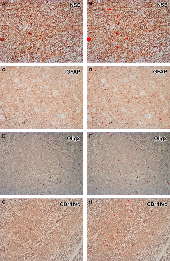
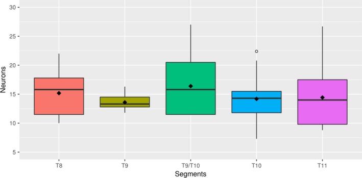
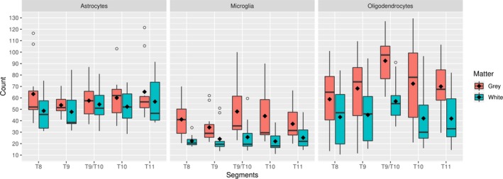
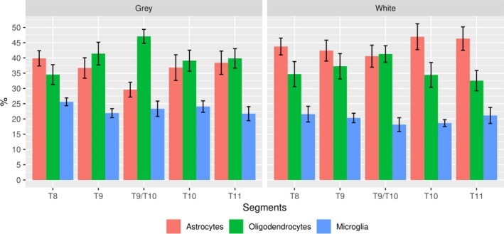
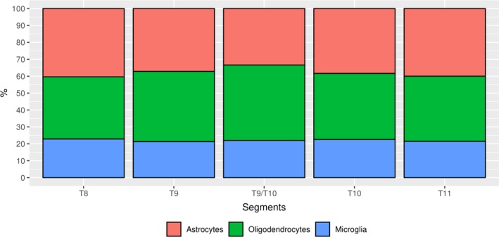
Spinal cord stimulation (SCS) applied between T8 and T11 segments has been shown to be effective for the treatment of chronic pain of the lower back and limbs. However, the mechanism of the analgesic effect at these medullary levels remains unclear. Numerous studies relate glial cells with development and maintenance of chronic neuropathic pain. Glial cells are electrically excitable, which makes them a potential therapeutic target using SCS. The aim of this study is to report glia to neuron ratio in thoracic segments relevant to SCS, as well as to characterize the glia cell population at these levels. Dissections from gray and white matter of posterior spinal cord segments (T8, T9, intersection T9/T10, T10 and T11) were obtained from 11 human cadavers for histological analyses. Neuronal bodies and glial cells (microglia, astrocytes and oligodendrocytes) were immunostained, microphotographed and counted using image analysis software. Statistical analyses were carried out to establish significant differences of neuronal and glial populations among the selected segments, between the glial cells in a segment, and glial cells in white and gray matter. Results show that glia to neuron ratio in the posterior gray matter of the human spinal cord within the T8-T11 vertebral region is in the range 11 : 1 to 13 : 1, although not significantly different among vertebral segments. Glia cells are more abundant in gray matter than in white matter, whereas astrocytes and oligodendrocytes are more abundant than microglia (40 : 40 : 20). Interestingly, the population of oligodendrocytes in the T9/T10 intersection is significantly larger than in any other segment. In conclusion, glial cells are the predominant bodies in the posterior gray and white matter of the T8-T11 segments of the human spinal cord. Given the crucial role of glial cells in the development and maintenance of neuropathic pain, and their electrophysiological characteristics, anatomical determination of the ratio of different cell populations in spinal segments commonly exposed to SCS is fundamental to understand fully the biological effects observed with this therapy.
Keywords: anatomy; glial cells; neurons; spinal cord.
© 2019 Anatomical Society.
Conflict of interest statement
The authors declare no conflicts of interest.
Figures
References
-
- Aarts E, Verhage M, Veenvliet JV, et al. (2014) A solution to dependency: using multilevel analysis to accommodate nested data. Nat Neurosci 17, 491–496. - PubMed
-
- Aló KM, Holsheimer J (2002) New trends in neuromodulation for the management of neuropathic pain. Neurosurgery 50, 690–703. - PubMed
-
- Azevedo FA, Carvalho LR, Grinberg LT, et al. (2009) Equal numbers of neuronal and nonneuronal cells make the human brain an isometrically scaled‐up primate brain. J Comp Neurol 513, 532–541. - PubMed
MeSH terms
LinkOut - more resources
Full Text Sources
Research Materials
