

Thalamic and cerebellar hypoperfusion in single photon emission computed tomography may differentiate multiple system atrophy and progressive supranuclear palsy
- PMID: 31348305
- PMCID: PMC6708712
- DOI: 10.1097/MD.0000000000016603
Thalamic and cerebellar hypoperfusion in single photon emission computed tomography may differentiate multiple system atrophy and progressive supranuclear palsy
Abstract
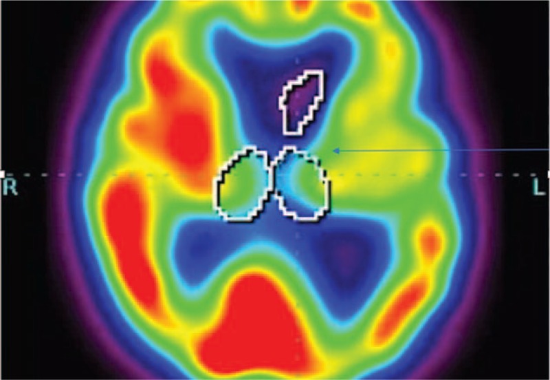
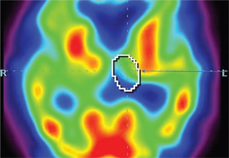
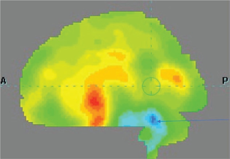
Neuroimaging in the context of examining atypical parkinsonian tauopathies is an evolving matter. Positron emission tomography and single photon emission computed tomography (SPECT) bring tools, which may be reasonable in supplementary examination, however, cannot be interpreted as a criterion standard for correct diagnosis. The aim of this observational study was to assess the differentiating potential of perfusion SPECT in 3 types of atypical parkinsonisms: multiple system atrophy parkinsonian type (MSA-P), corticobasal syndrome (CBS), and progressive supranuclear palsy (PSP). The study was carried out using the comparison of standard deviations of perfusion in patients from these 3 groups. Data obtained from 10 patients with clinical diagnosis MSA-P, 14 patients with CBS and 21 patients with PSP, which were analyzed using Tukey honest significant difference post-hoc test, revealed significant differences of perfusion P < .05 between MSA-P and PSP within the cerebellum and thalamus. No significant differences between CBS and PSP were observed.
Conflict of interest statement
The authors have no conflicts of interest to disclose.
Figures
References
-
- Valotassiou V, Papatriantafyllou J, Sifakis N, et al. Brain perfusion SPECT with Brodmann areas analysis in differentiating frontotemporal dementia subtypes. Curr Alzheimer Res 2014;11:941–54. - PubMed
-
- Sławek J, Lass P, Derejko M, et al. Cerebral blood flow SPECT may be helpful in establishing the diagnosis of progressive supranuclear palsy and corticobasal degeneration. Nucl Med Rev Cent East Eur 2001;4:73–6. - PubMed
Publication types
MeSH terms
LinkOut - more resources
Full Text Sources
Medical
Miscellaneous
