Review
. 2019 Nov 21;28(R2):R219-R225.
doi: 10.1093/hmg/ddz178.

Synapse diversity and synaptome architecture in human genetic disorders

Affiliations
Review

Synapse diversity and synaptome architecture in human genetic disorders

Seth G N Grant. Hum Mol Genet. .

Abstract

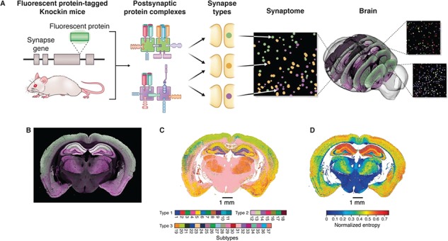
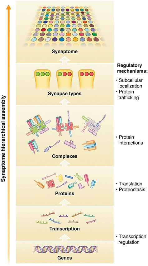
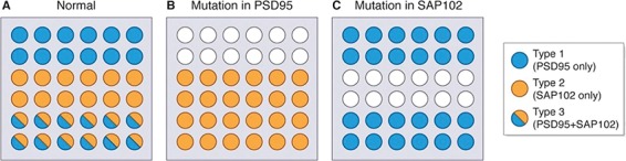
Over 130 brain diseases are caused by mutations that disrupt genes encoding the proteome of excitatory synapses. These include neurological and psychiatric disorders with early and late onset such as autism, schizophrenia and depression and many other rarer conditions. The proteome of synapses is highly complex with over 1000 conserved proteins which are differentially expressed generating a vast, potentially unlimited, number of synapse types. The diversity of synapses and their location in the brain are described by the synaptome. A recent study has mapped the synaptome across the mouse brain, revealing that synapse diversity is distributed into an anatomical architecture observed at scales from individual dendrites to the whole systems level. The synaptome architecture is built from the hierarchical expression and assembly of proteins into complexes and supercomplexes which are distributed into different synapses. Mutations in synapse proteins change the synaptome architecture leading to behavioral phenotypes. Mutations in the mechanisms regulating the hierarchical assembly of the synaptome, including transcription and proteostasis, may also change synapse diversity and synaptome architecture. The logic of synaptome hierarchical assembly provides a mechanistic framework that explains how diverse genetic disorders can converge on synapses in different brain circuits to produce behavioral phenotypes.


Figures

Figure 1
Figure 1
Synaptome mapping in mouse. Genes expressing synapse proteins are tagged in mice by fusing a genetically encoded fluorescent protein onto the C-terminus of postsynaptic scaffold proteins that assemble postsynaptic signaling complexes (PSD95, green; SAP102, magenta). These complexes are distributed into different synapse types that can be visualized with confocal microscopy. The synaptome map is built by quantification of synapse types from regions of the mouse brain. Example image of a coronal mouse brain section showing the differential distribution of PSD95 (green) and SAP102 (magenta). A synaptome map of a coronal section showing the dominant or major subtype from 37 subtypes in different regions. A synaptome map showing the extent of synapse diversity in different regions of the mouse brain. Figures adapted from Zhu et al., 2018 (31).
Figure 2
Figure 2
Synaptome hierarchical assembly. The diversity of synapse types and their spatial distribution in the synaptome arise from a hierarchical regulatory mechanism controlling gene and protein expression, assembly of proteins into complexes and supercomplexes and distribution of these supramolecular assemblies into synapses. Mutations acting on regulatory mechanisms at all levels of the hierarchy could influence synapse diversity and synaptome architecture.
Figure 3
Figure 3
Mutations reprogram synaptome architecture. Model of a normal synaptome comprised of 36 synapses made of three types assembled from two proteins (PSD95 and SAP102). Type 1 synapses express only PSD95, type 2 expresses only SAP102 and type 3 a mixture of both proteins. A mutation that knocks out PSD95 changes the synaptome architecture by abolishing type 1 synapses (empty circles in top two rows) and convert the type 3 synapses to type 2 synapses. A mutation that knocks out SAP102 does not affect type 1 synapses, but abolishes type 2 synapses and converts type 3 synapses into type 2 synapses.

Similar articles

  • Architecture of the Mouse Brain Synaptome.
    Zhu F, Cizeron M, Qiu Z, Benavides-Piccione R, Kopanitsa MV, Skene NG, Koniaris B, DeFelipe J, Fransén E, Komiyama NH, Grant SGN. Zhu F, et al. Neuron. 2018 Aug 22;99(4):781-799.e10. doi: 10.1016/j.neuron.2018.07.007. Epub 2018 Aug 2. Neuron. 2018. PMID: 30078578 Free PMC article.
  • Hierarchical organization and genetically separable subfamilies of PSD95 postsynaptic supercomplexes.
    Frank RAW, Zhu F, Komiyama NH, Grant SGN. Frank RAW, et al. J Neurochem. 2017 Aug;142(4):504-511. doi: 10.1111/jnc.14056. Epub 2017 Jul 25. J Neurochem. 2017. PMID: 28452394 Free PMC article.
  • A brain atlas of synapse protein lifetime across the mouse lifespan.
    Bulovaite E, Qiu Z, Kratschke M, Zgraj A, Fricker DG, Tuck EJ, Gokhale R, Koniaris B, Jami SA, Merino-Serrais P, Husi E, Mendive-Tapia L, Vendrell M, O'Dell TJ, DeFelipe J, Komiyama NH, Holtmaat A, Fransén E, Grant SGN. Bulovaite E, et al. Neuron. 2022 Dec 21;110(24):4057-4073.e8. doi: 10.1016/j.neuron.2022.09.009. Epub 2022 Oct 5. Neuron. 2022. PMID: 36202095 Free PMC article.
  • Synaptopathies: diseases of the synaptome.
    Grant SG. Grant SG. Curr Opin Neurobiol. 2012 Jun;22(3):522-9. doi: 10.1016/j.conb.2012.02.002. Epub 2012 Mar 10. Curr Opin Neurobiol. 2012. PMID: 22409856 Review.
  • The postsynaptic organization of synapses.
    Sheng M, Kim E. Sheng M, et al. Cold Spring Harb Perspect Biol. 2011 Dec 1;3(12):a005678. doi: 10.1101/cshperspect.a005678. Cold Spring Harb Perspect Biol. 2011. PMID: 22046028 Free PMC article. Review.

Cited by

References

    1. Husi H., Ward M.A., Choudhary J.S., Blackstock W.P. and Grant S.G. (2000) Proteomic analysis of NMDA receptor-adhesion protein signaling complexes. Nat Neurosci., 3, 661–669. - PubMed
    1. Husi H. and Grant S.G. (2001) Isolation of 2000-kDa complexes of N-methyl-D-aspartate receptor and postsynaptic density 95 from mouse brain. J. Neurochem., 77, 281–291. - PubMed
    1. Bayes A., Collins M.O., Croning M.D., van de Lagemaat L.N., Choudhary J.S. and Grant S.G. (2012) Comparative study of human and mouse postsynaptic proteomes finds high compositional conservation and abundance differences for key synaptic proteins. PLoS One, 7, e46683. - PMC - PubMed
    1. Bayes A., Collins M.O., Reig-Viader R., Gou G., Goulding D., Izquierdo A., Choudhary J.S., Emes R.D. and Grant S.G. (2017) Zebrafish synapse proteome complexity, evolution and ultrastructure. Nat. Commun., 8, 14613. - PMC - PubMed
    1. Bayes A., van de Lagemaat L.N., Collins M.O., Croning M.D., Whittle I.R., Choudhary J.S. and Grant S.G. (2011) Characterization of the proteome, diseases and evolution of the human postsynaptic density. Nat. Neurosci., 14, 19–21. - PMC - PubMed

Publication types

MeSH terms