. 2019 Jul 26;15(7):e1007946.
doi: 10.1371/journal.ppat.1007946. eCollection 2019 Jul.

Toxoplasma F-box protein 1 is required for daughter cell scaffold function during parasite replication

Affiliations

Toxoplasma F-box protein 1 is required for daughter cell scaffold function during parasite replication

Carlos Gustavo Baptista et al. PLoS Pathog. .

Abstract

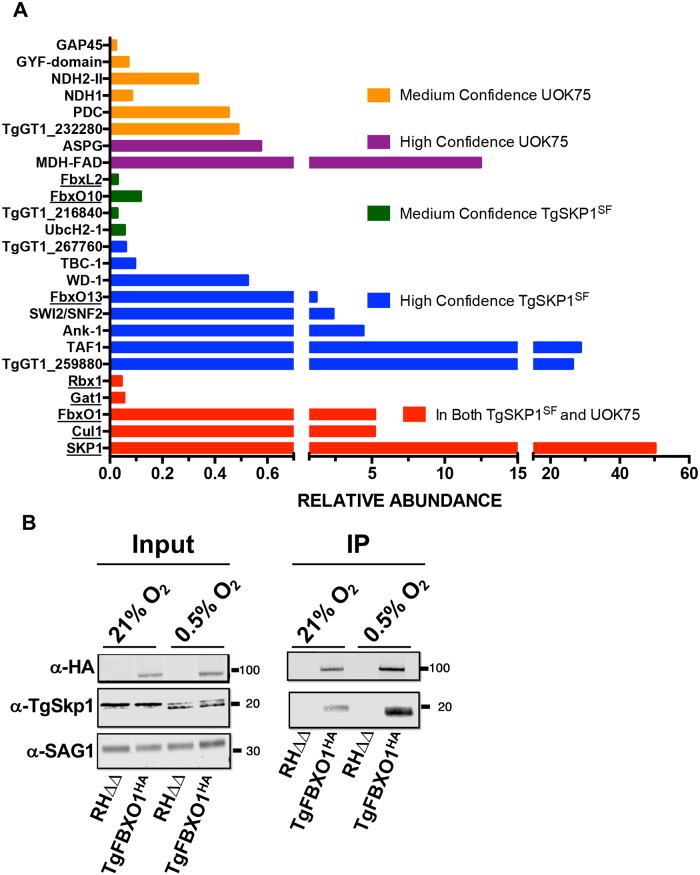
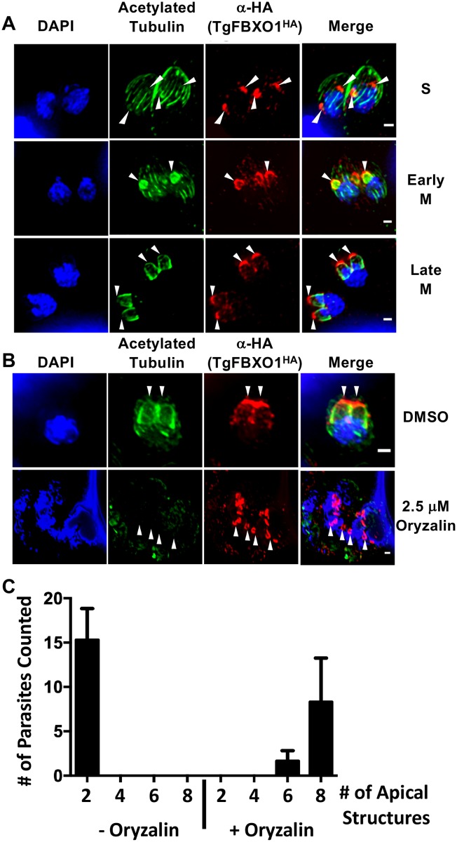
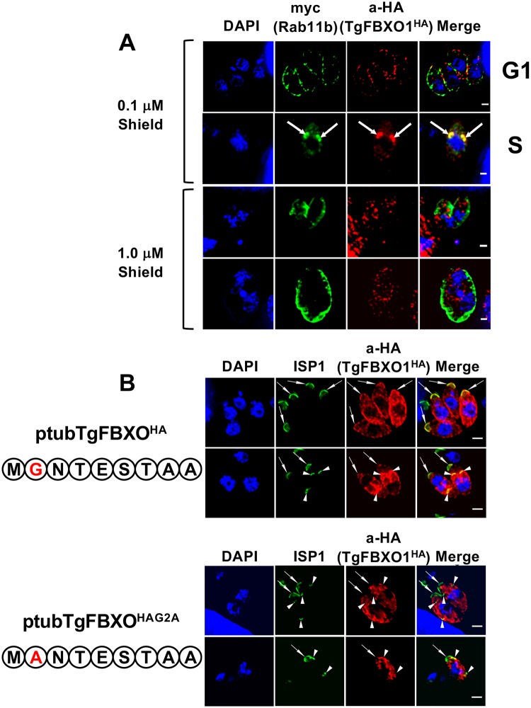
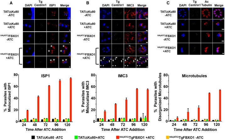
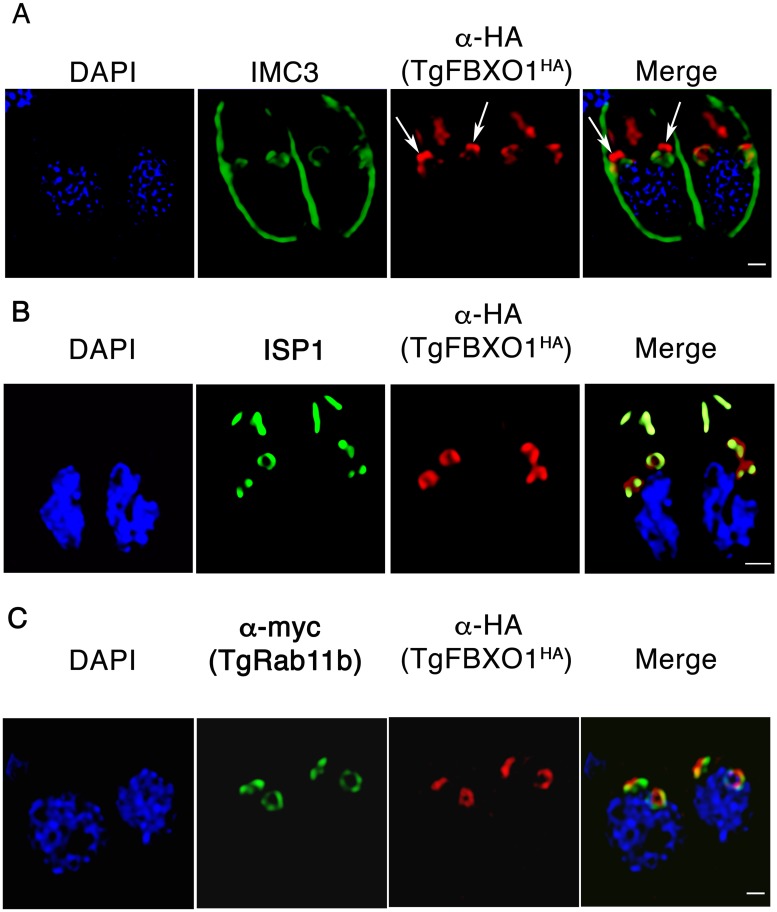
By binding to the adaptor protein SKP1 and serving as substrate receptors for the SKP1 Cullin, F-box E3 ubiquitin ligase complex, F-box proteins regulate critical cellular processes including cell cycle progression and membrane trafficking. While F-box proteins are conserved throughout eukaryotes and are well studied in yeast, plants, and animals, studies in parasitic protozoa are lagging. We have identified eighteen putative F-box proteins in the Toxoplasma genome of which four have predicted homologs in Plasmodium. Two of the conserved F-box proteins were demonstrated to be important for Toxoplasma fitness and here we focus on an F-box protein, named TgFBXO1, because it is the most highly expressed by replicative tachyzoites and was also identified in an interactome screen as a Toxoplasma SKP1 binding protein. TgFBXO1 interacts with Toxoplasma SKP1 confirming it as a bona fide F-box protein. In interphase parasites, TgFBXO1 is a component of the Inner Membrane Complex (IMC), which is an organelle that underlies the plasma membrane. Early during replication, TgFBXO1 localizes to the developing daughter cell scaffold, which is the site where the daughter cell IMC and microtubules form and extend from. TgFBXO1 localization to the daughter cell scaffold required centrosome duplication but before kinetochore separation was completed. Daughter cell scaffold localization required TgFBXO1 N-myristoylation and was dependent on the small molecular weight GTPase, TgRab11b. Finally, we demonstrate that TgFBXO1 is required for parasite growth due to its function as a daughter cell scaffold effector. TgFBXO1 is the first F-box protein to be studied in apicomplexan parasites and represents the first protein demonstrated to be important for daughter cell scaffold function.


Conflict of interest statement

The authors have declared that no competing interests exist.

Figures

Fig 1
Fig 1. TgFBXO1 interacts with TgSKP1.
(A). RHΔhxgprtΔKu80 and TgSKP1SF tachyzoites were solubilized and immunoprecipitated with affinity purified anti-TgSKP1 (UOK75) or anti-FLAG (M2), respectively. The immunoprecipitates were trypsin digested and subjected to proteomic analysis. Protein candidates were selected from a list of protein hits that were detected in multiple replicates and minimally recovered with irrelevant antibodies (for UOK75) or the untagged strain (for M2), and are reported as found in either both (red bars) or only individual immunoprecipitations with either high or medium confidence. Proteins known or predicted to interact with TgSKP1 are underlined. See S2 Fig and S2 Table for origin of protein labels and more information about control data. (B). Lysates prepared from TgFBXO1HA and parental RHΔhxgprtΔKu80 grown at 21% or 0.5% O2 were either Western blotted (Input) or incubated with anti-HA antibodies (IP). The immunoprecipitates were captured by Protein G Sepharose and the immune complexes were Western blotted to detect TgFBXO1HA, TgSkp1, and SAG1.
Fig 2
Fig 2. TgFBXO1 is important for Toxoplasma growth.
(A). Schematic illustration of HA(ATC)TgFBXO1 anhydrotetracycline (ATC)-regulated gene expression. The TgFBXO1 endogenous promoter was replaced with a SAG4 promoter containing a tetracycline transactivator (tTA) binding element. In the absence of ATC, the tetracycline regulated promoter is active, while the addition of ATC reduces transcription by preventing tTA from binding to and activating the promoter. (B). Lysates from HA(ATC)TgFBXO1 or parental TATiΔKu80 parasites grown for 24 h in the absence or presence of 1μg/ml ATC were Western blotted to detect HA(ATC)TgFBXO1 or SAG1 as a loading control. (C). HA(ATC)TgFBXO1 or TATiΔKu80 parasites (100/well of a 6 well plate) were grown for 5 d on HFF monolayers with or without 1μg/ml ATC. The cells were then fixed and numbers of plaques formed counted. Shown are the averages and standard deviations of 3 independent experiments performed in triplicate. (D). Representative images of plaques from (C) showing decreased plaque size of TgFBXO1-depleted parasites. Plaque sizes were quantified and plotted on the adjacent graph. *, p < 0.001, One-Way ANOVA. Bars = 0.5mm.
Fig 3
Fig 3. TgFBXO1 forms apical structures early during endodyogeny.
(A). TgFBXO1HA-expressing parasites were fixed and stained to detect IMC3, TgFBXO1, and DNA during G1, S, and M phases. Arrows highlight TgFBXO1 apical structures. (B). TgFBXO1HA-expressing parasites were fixed and stained to detect ISP1, TgFBXO1, and DNA during S, early M (before nuclear segregation), and late M (during nuclear segregation) phases. Arrows highlight TgFBXO1 apical structures. Bars = 2μm. (C). TOP: Fractionation scheme to characterize TgFBXO1HA association with the IMC. BOTTOM: Western blotting of equivalent volumes of each indicated fraction. Blots were probed with antibodies to detect TgFBXO1 (α-HA), IMC3 (αIMC3), and ISP1 (αISP1).
Fig 4
Fig 4. TgFBXO1 apical structures form after centrosome duplication and are localized apically to the parasite centrosome.
(A). TgFBXO1HA-expressing parasites were fixed and stained to detect TgCentrin1, TgFBXO1, and DNA during S, early M (EM), and late M (LM) phases. Arrowheads highlight TgFBXO1 apical structures and arrows highlight TgCentrin1. Bars = 2μm. A schematic depicting the cell cycle phases is shown. (B). Parasites were treated with 3 μM SB505124 (or DMSO as a vehicle control) for 24 h, fixed and then stained to detect TgCentrin1, TgFBXO1, and DNA. Note the appearance of SB505124-treated parasites with >2 centrin1+ foci and disorganized TgFBXO1 staining. Bars = 5 μm. (C). Quantification of numbers of parasites with apical TgFBXO1 structures. Data represents averages and standard deviations of three independent experiments with at least 50 parasites examined/experiment (D). TgFBXO1HA parasites were stained to detect TgFBXO1 and the kinetochore marker TgNuf2. Note TgFBXO1HA apical structures form after kinetochore duplication but before separation is completed as evidenced by the lobed TgNuf2 staining (Insert). Bars: 2μm.
Fig 5
Fig 5. TgFBXO1 apical structures form independently of daughter cell microtubule assembly.
(A). TgFBXO1HA-expressing parasites were stained to detect TgFBXO1, acetylated tubulin (to detect newly synthesized microtubules), and DNA during S, early M, and late M (during nuclear segregation) phases. Bars = 2 μm. (B). Parasites were treated with oryzalin (2.5 μM) or DMSO (vehicle control) for 24 h and then stained to detect TgFBXO1, acetylated tubulin, and DNA. Arrowheads highlight TgFBXO1 apical structures. Bars = 2 μm. (C). Quantification of numbers of TgFBXO1HA structures per parasite that form in the presence of DMSO or oryzalin. Shown are the averages and standard deviations of 3 independent experiments in which 50 randomly selected parasites were counted.
Fig 6
Fig 6. TgFBXO1 is recruited to the DCS in a Rab11b-dependent manner.
(A). TgFBXO1HA-expressing parasites were transiently transfected with p5RT70DDmycRab11b-HXGPRT expression construct and then grown in the presence of Shield-1 (0.1μM—low level expression or 1 μM–high level expression) reagent for 48 h. Cells were then fixed and stained to detect TgRab11b or TgFBXO1. Arrows highlight apparent colocalization of TgRab11b with the TgFBXO1 apical structures. Note peripheral TgRab11b and punctate TgFBXO1 localization in parasites treated with 1 μM Shield-1. Bars = 2μm. (B). Parasites were transiently transfected with wild-type (ptubTgFBXO1HA) or myristoylation mutant (ptubTgFBXO1HAG2A) TgFBXO1 expression constructs, fixed 48 h later, and stained to detect ISP1 and TgFBXO1. Arrows highlight position of ISP1 apical caps and arrowheads highlight DCS structures. Bars = 2 μm.
Fig 7
Fig 7. TgFBXO1 is required for IMC organization and maturation.
(A-C). Parental RHΔhxgprtΔKu80 and HA(ATC)TgFBXO1 parasites were grown for 1–5 days in the absence or presence of ATC (1 μg/ml) and stained to detect DNA (blue), centrin (green), and ISP1, IMC3, or acetylated tubulin (red). Images are of representative parasites 5 days -/+ ATC. Graphs below each panel represent averages and standard deviations from 3 independent experiments with at least 50 parasites counted per condition. Bars = 2 μm. (A). * denotes mislocalized ISP1 in DCS of parasites with decreased TgFBXO1 expression. Bars = 1 μm. (B). ⨁ highlights IMC3 whirls, * highlights parasites lacking IMC3 staining, arrows highlight misaligned IMC3 and centrin1, and arrowheads highlight parasites lacking centrin1 staining. Bars = 2 μm. (C). Note disorganized appearance of acetylated microtubules in ATC-treated HA(ATC)TgFBXO1 parasites. Bars = 2 μm.
Fig 8
Fig 8. Super resolution microscopy imaging of TgFBXO1.
TgFBXO1HA parasites were fixed and imaged using Hyvolution Acquisition Software to detect TgFBXO1 and IMC3 (A), ISP1 (B), and TgRab11b (C). Shown are still images from movies of three dimensional projections available as supplemental data.

Similar articles

Cited by

References

    1. Guo M, Mishra A, Buchanan RL, Dubey JP, Hill DE, Gamble HR, et al. A Systematic Meta-Analysis of Toxoplasma gondii Prevalence in Food Animals in the United States. Foodborne Pathog Dis. 2016;13(3):109–18. 10.1089/fpd.2015.2070 . - DOI - PubMed
    1. Pappas G, Roussos N, Falagas ME. Toxoplasmosis snapshots: global status of Toxoplasma gondii seroprevalence and implications for pregnancy and congenital toxoplasmosis. Int J Parasitol. 2009;39(12):1385–94. Epub 2009/05/13. 10.1016/j.ijpara.2009.04.003 . - DOI - PubMed
    1. Dubey JP. History of the discovery of the life cycle of Toxoplasma gondii. Int J Parasitol. 2009;39(8):877–82. Epub 2009/07/25. . - PubMed
    1. Bierly AL, Shufesky WJ, Sukhumavasi W, Morelli AE, Denkers EY. Dendritic cells expressing plasmacytoid marker PDCA-1 are Trojan horses during Toxoplasma gondii infection. J Immunol. 2008;181(12):8485–91. Epub 2008/12/04. 10.4049/jimmunol.181.12.8485 - DOI - PMC - PubMed
    1. Weiss LM, Kim K. The development and biology of bradyzoites of Toxoplasma gondii. Front Biosci. 2000;5:D391–405. . - PMC - PubMed

Publication types

MeSH terms