. 2019 Oct:21:102067.
doi: 10.1016/j.nano.2019.102067. Epub 2019 Jul 23.

Towards a nanoparticle-based prophylactic for maternal autoantibody-related autism

Affiliations

Towards a nanoparticle-based prophylactic for maternal autoantibody-related autism

Amir Bolandparvaz et al. Nanomedicine. 2019 Oct.

Abstract

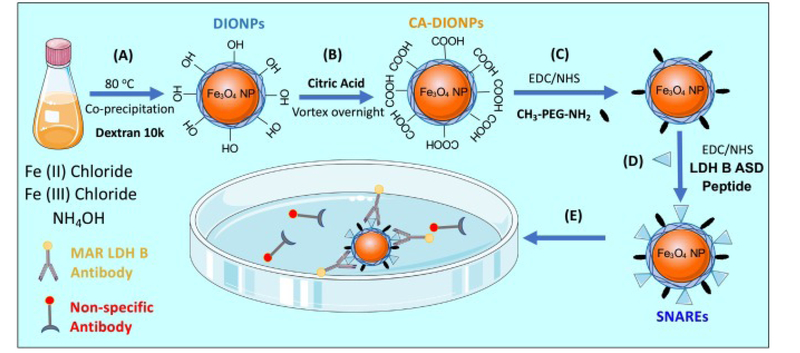
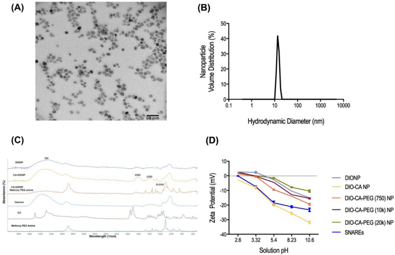
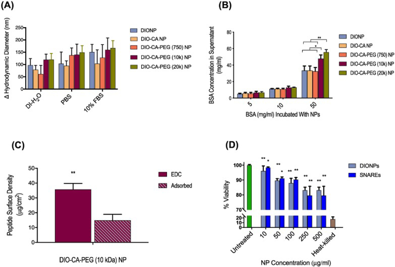
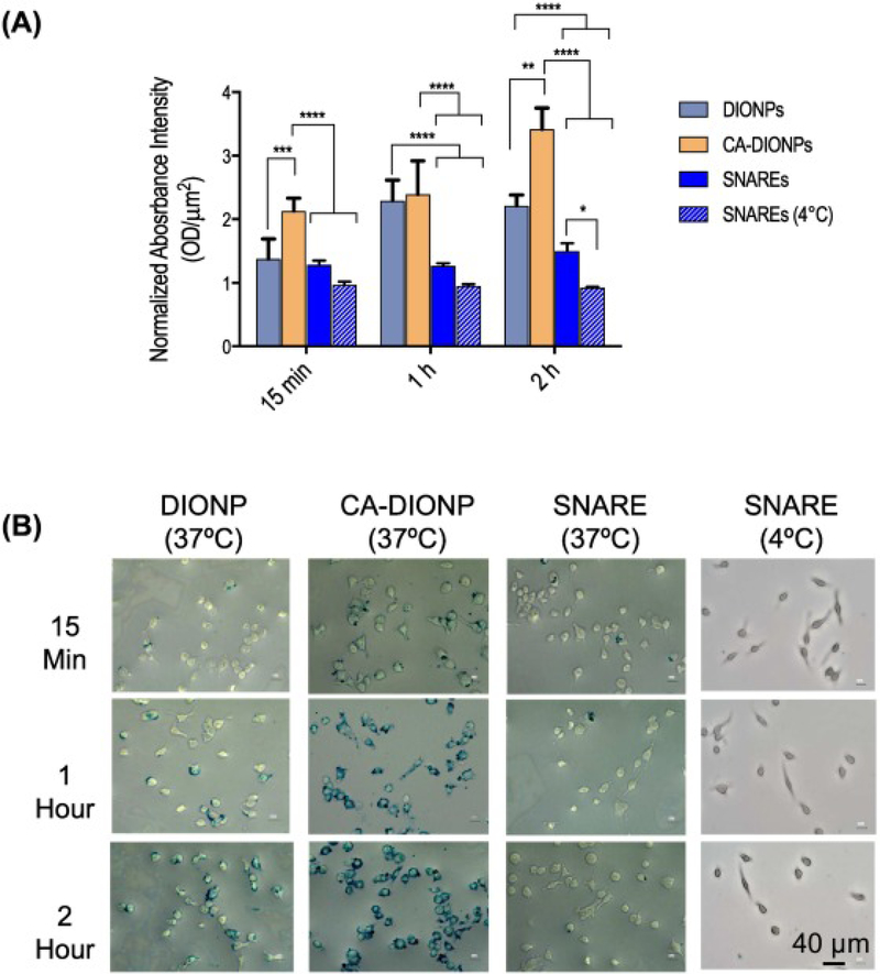
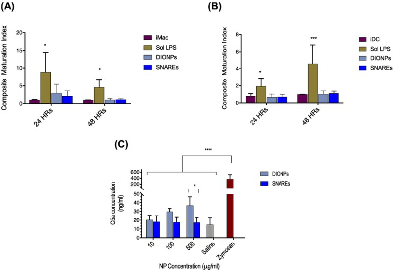
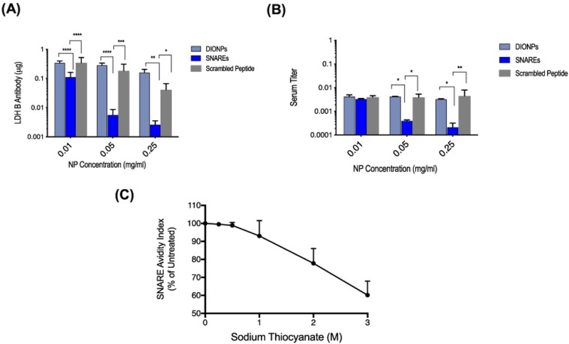
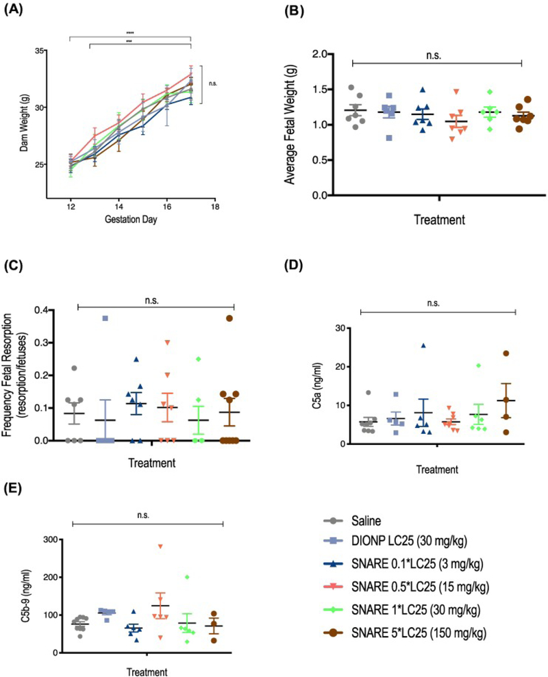
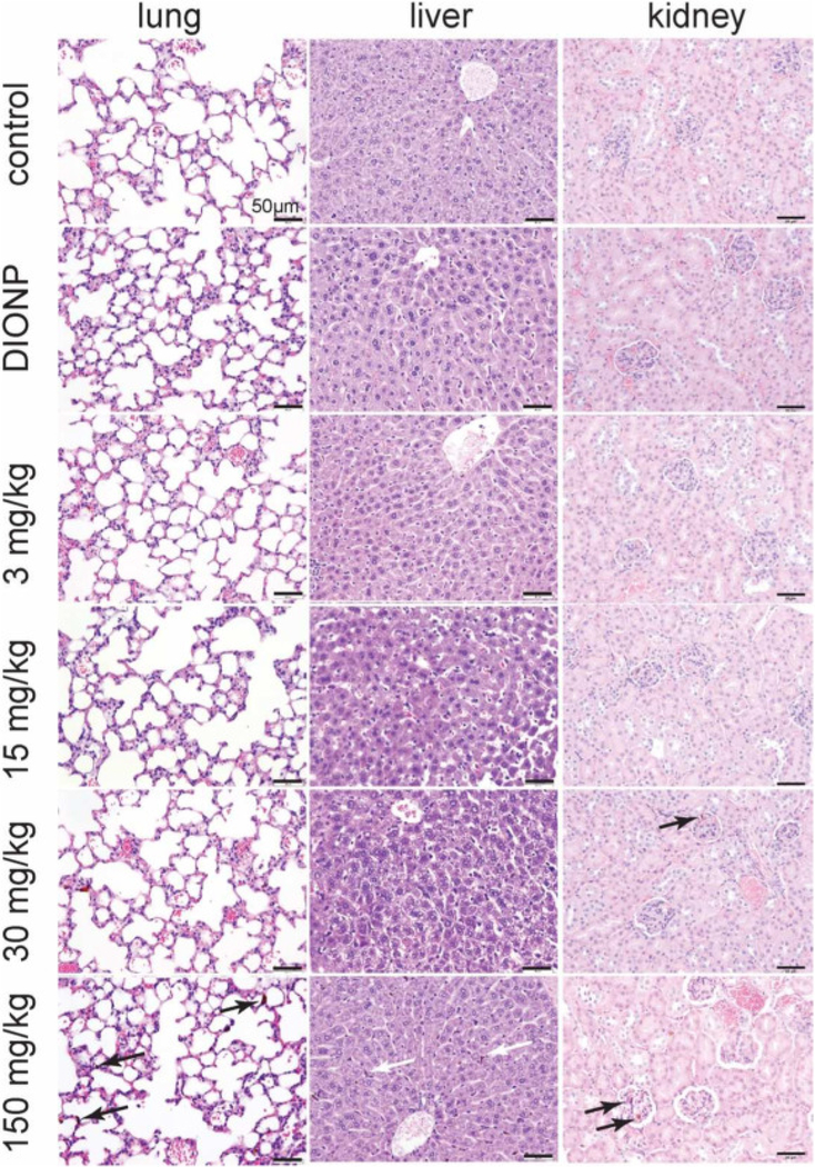
Recently, the causative agents of Maternal Autoantibody-Related (MAR) autism, pathological autoantibodies and their epitopic targets (e.g. lactate dehydrogenase B [LDH B] peptide), have been identified. Herein, we report on the development of Systems for Nanoparticle-based Autoantibody Reception and Entrapment (SNAREs), which we hypothesized could scavenge disease-propagating MAR autoantibodies from the maternal blood. To demonstrate this functionality, we synthesized 15 nm dextran iron oxide nanoparticles surface-modified with citric acid, methoxy PEG(10 kDa) amine, and LDH B peptide (33.8 μg peptide/cm2). In vitro, we demonstrated significantly lower macrophage uptake for SNAREs compared to control NPs. The hallmark result of this study was the efficacy of the SNAREs to remove 90% of LDH B autoantibody from patient-derived serum. Further, in vitro cytotoxicity testing and a maximal tolerated dose study in mice demonstrated the safety of the SNARE formulation. This work establishes the feasibility of SNAREs as the first-ever prophylactic against MAR autism.

Keywords: Iron oxide(2); Maternal autoantibody-related autism(1); Peptide-functionalized(4); nanoparticles(3).


Conflict of interest statement

Conflict of Interest: Dr. Van de Water has a patent application involving the MAR ASD peptides described herein; all other authors have no conflicts of interest to declare.

Figures

Figure 1.
Figure 1.
Schematic summarizing synthesis of peptide-functionalized dextran iron oxide nanoparticles and capture of Autism-specific autoantibodies from an antibody solution. (A) Synthesis of dextran iron oxide nanoparticles (DIONPS) via co-precipitation method. (B) Exchange of DIONP surface hydroxyl groups with carboxylic groups by citric acid coating (DIO-CA NPs). (C) Nanoparticle surface-conjugation with Methoxy PEG Amine via EDC/NHS reaction (DIO-CA-PEG NPs). (D) Nanoparticle surface-decoration with LDH B ASD peptide via EDC/NHS Reaction (SNAREs). (E) SNAREs specifically ligate Anti-LDH B autoantibody from solution.
Figure 2.
Figure 2.
SNARE physico-chemical characterization. (A) Transmission electron microscopy (TEM) of DIONPs. (B) Dynamic light scattering (DLS). (C) FTIR spectra of DIONPs, citric acid DIONPs, DIO-CA-PEG (10 kDa) NPs. (D) The zeta potential for naked DIONPs, DIO-CA NPs, and methoxy PEG amine (750 Da, 10 kDa, or 20 kDa)-coated DIO-CA NPs (1 mg/ml).
Figure 3.
Figure 3.
Nanoparticle in vitro characterization and MAR peptide conjugation. (A) No significant nanoparticle aggregation was observed in various media (DI-H2O, PBS, or 10% FBS in PBS) after 24 h incubation (measured using DLS). (B) PEG (10 kDa/ 20 kDa)-conjugated DIO-CA NPs had the highest repulsion ability, preventing adsorption of non-specific proteins (Bovine Serum Albumin [BSA]). BSA adsorption on DIONP formulations normalized to the number of DIONPs per batch. (C) Conjugation of FITC-LDH B peptide onto DIO-CA-PEG (10 kDa) NPs resulted in a final surface density of 33.8 μg peptide/cm2. (D) In vitro, the LC25 for RAW 264.7 macrophage cells incubated with DIONPs and SNAREs was determined to be 500 μg/ml. Heat-killed and untreated cells were used as control and treatments normalized to untreated cells.
Figure 4.
Figure 4.
Nanoparticle uptake by macrophages. (A) In vitro uptake of DIONPs, DIO-CA NPs, ad SNAREs by RAW 264.7 macrophages was assessed using a Prussian Blue iron stain after 15 min, 1 h, and 2 h of incubation at either 4 or 37 °C. (B) Representative images of Prussian Blue staining.
Figure 5.
Figure 5.
In vitro assessment of complement activation–C5a generation and phenotypic analysis of dendritic cells and macrophages. (A) Bone marrow-derived macrophages demonstrated no significant change in maturation when treated with SNAREs or DIONPs, compared to immature macrophages. (B) Bone-marrow-derived DC maturation is unaffected by the presence of DIONPS or SNAREs. (C) C5a complement fragment concentration in serum of C57BL/6j mice incubated with DIONPs, SNAREs, saline control, and zymosan positive control for 4 h at 37 °C.
Figure 6.
Figure 6.
Assessment of MAR autoantibody capture, specificity, and affinity in vitro. (A) SNAREs captured up to 2.7 μg LDH B Ab/ mg SNAREs (95% efficiency) and exhibited increased antibody entrapment at higher concentrations. (B) SNAREs reduced LDH B antibody titer in human serum by as much as 90% (patient-derived serum of mothers of children with ASD). Scrambled peptide-DIONPs and DIONPs were used as negative controls. (C) Avidity of SNAREs to MAR LDH B antibody indicating a 50% avidity index at ~3.6 M sodium thiocyanate concentration.
Figure 7.
Figure 7.
SNARE maximum tolerated dose (MTD) and assessment of complement activation in pregnant dams. (A) Administration of SNAREs did not affect prenatal weight gain. Mice were weighted post-injection from GD 12 to GD 17, when mice were sacrificed for post-mortem analysis. (B) Average fetal weight (AFW), determined by dividing the total weight of the uteri by the number of non-resorbed fetuses, is shown. (C) Frequency of fetal resorption calculated as number of resorption/number of fetuses of treated compared to saline-treated mice. (D) C5a complement factor concentration in serum of mice treated with SNAREs showed no significant differences to the saline control. (E) C5b-9 complement factor concentration in serum of SNARE-treated mice also demonstrated no significant differences compared to saline control.
Figure 8.
Figure 8.
Histological assessment of SNAREs in lungs, liver, and kidney of pregnant dams. Minimal histological changes were evident in pregnant dams intravenously injected with 3 or 15 mg NPs/kg of SNAREs on GD 12 and sacrificed on GD 17. At 30 mg NPs/kg injection dose of SNAREs and DIONPs, brown pigment (iron deposits) was present in Kupffer cells and rarely within pulmonary capillaries with no evidence of hepatic or lung damage. At the maximal dose of 150 mg/kg, arrows point to high amounts of intravascular iron aggregates observed in the lung sections with mild degree of congestion. In the liver, brown pigment was observed at moderate to high frequency within Kupffer cells. In the kidney, the brown pigment was present with in glomerular capillaries and occasionally in the renal venules. No evidence of tissue damage or inflammation was associated with the presence of the intravascular deposits in the lung or intracellular pigment in the liver.

Similar articles

Cited by

References

    1. DSM-5 autism spectrum disorder fact sheet. Association AP. 2013.
    1. Christensen DL, Baio J, Van Naarden Braun K., Bilder D, Charles J, Constantino JN, et al.Prevalence and characteristics of autism Spectrum disorder among children aged 8 years—autism and developmental disabilities monitoring network, 11 sites, United States, 2012 MMWR Surveill Summ, 65 (3) (2016), pp. 1–23 - PMC - PubMed
    1. Leigh JP, Du J. Brief report: forecasting the economic burden of autism in 2015 and 2025 in the United States J Autism Dev Disord, 45 (12) (2015), pp. 4135–4139 - PubMed
    1. DeFilippis M, Wagner KD Treatment of autism spectrum disorder in children and adolescents Psychopharmacol Bull, 46 (2) (2016), pp. 18–41 - PMC - PubMed
    1. Onore C, Careaga M, Ashwood P. The role of immune dysfunction in the pathophysiology of autism Brain Behav Immun, 26 (3) (2012), pp. 383–392 - PMC - PubMed

Publication types

MeSH terms