. 2019 Jul 25;9(8):1069.
doi: 10.3390/nano9081069.

Carbon Nano-Onions as Non-Cytotoxic Carriers for Cellular Uptake of Glycopeptides and Proteins

Affiliations

Carbon Nano-Onions as Non-Cytotoxic Carriers for Cellular Uptake of Glycopeptides and Proteins

Marta d'Amora et al. Nanomaterials (Basel). .

Abstract

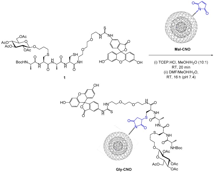
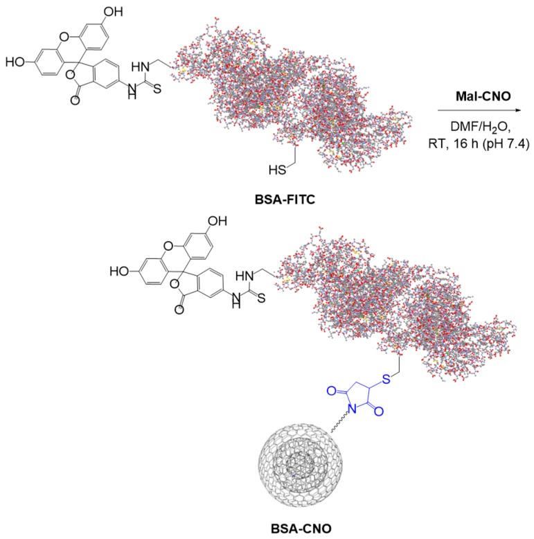
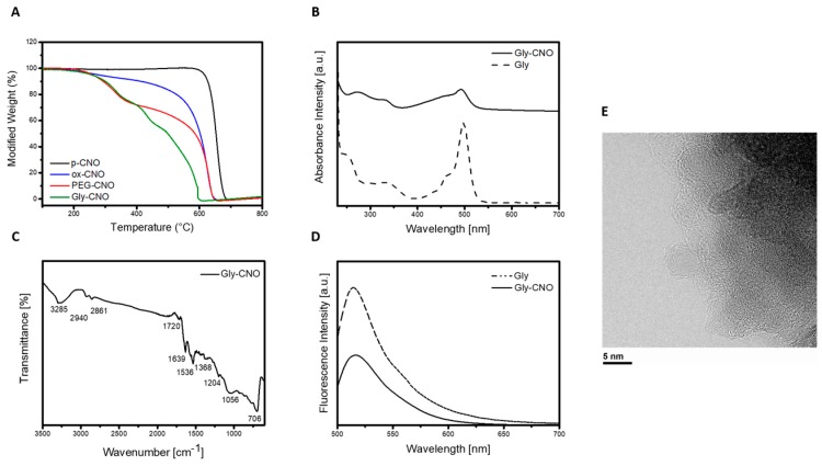
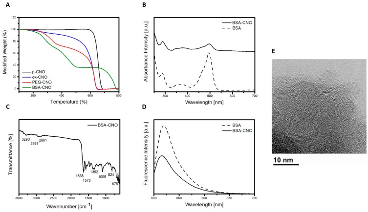
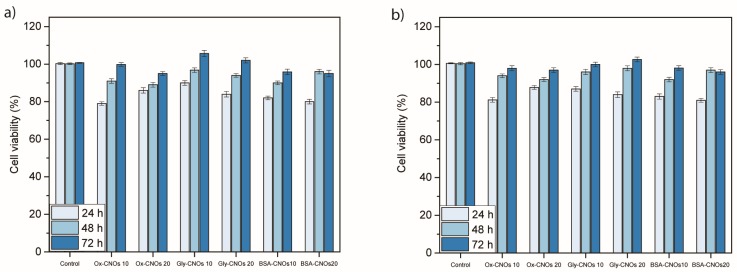
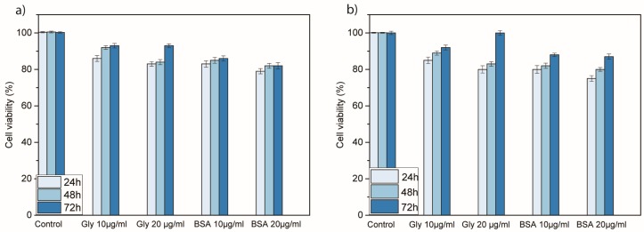
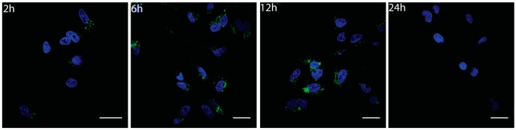
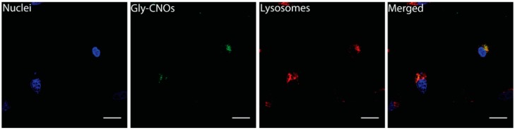
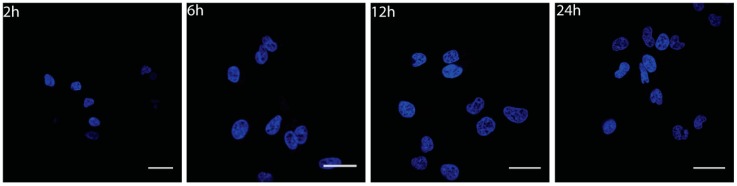
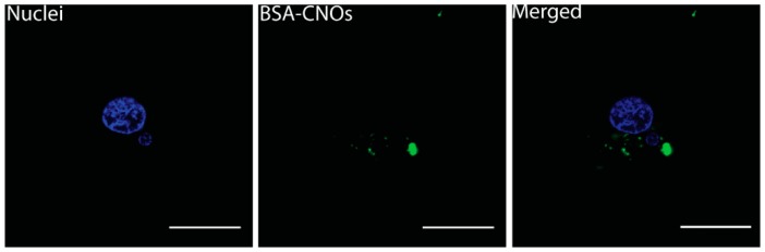
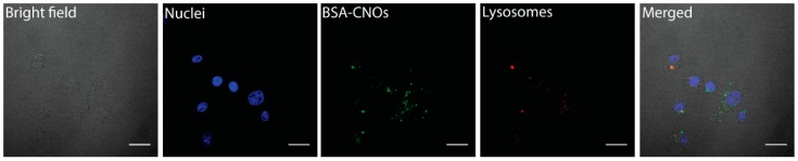
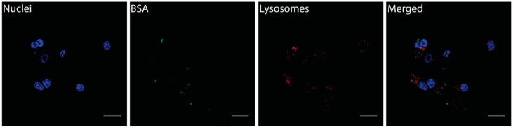
Carbon nano-onions (CNOs) possess favorable properties that make them suitable for biomedical applications, including their small size, ready surface modification, and good biocompatibility. Here, we report the covalent immobilization of a synthetic glycopeptide and the protein bovine serum albumin (BSA) onto the surface of carbon nano-onions using the maleimide-thiol "addition reaction". The glycopeptide and BSA are readily transported inside different cell lines, together with carbon nano-onions, through the endocytosis pathway. Our results show that carbon nano-onions are excellent scaffolds for glycopeptides and proteins immobilization and act as intracellular carriers for these biomolecules. These findings open new perspectives in the application of carbon nano-onions as intracellular transporters in diverse biomedical applications.

Keywords: biomedical applications; biomolecules; carbon nano-onions; carriers; scaffolds.


Conflict of interest statement

The authors declare no conflict of interest.

Figures

Scheme 1
Scheme 1
Ligation reaction between synthetic glycopeptide 1 and maleimido decorated CNO (Mal-CNO).
Scheme 2
Scheme 2
Ligation reaction between BSA-FITC and Mal-CNO.
Figure 1
Figure 1
Characterization of the functionalized glycopeptide-CNO conjugate (Gly-CNO). (A) Thermogravimetric analysis of p-CNO (black), ox-CNO (blue), PEG-CNO (red), and Gly-CNO (green). All experiments were run in air with a temperature rate of 10 °C min−1. (B) Absorption spectra of Gly-CNO (solid line) and fluorescent labeled glycopeptide 1 (dotted line) in DMEM phenol red free at a concentration of 10 μg mL−1. (C) ATR-FTIR spectra of Gly-CNO using germanium crystals and 256 scans. (D) Emission spectra (λexc = 490 nm) of fluorescent labeled glycopeptide 1 (dotted line) and Gly-CNO (solid line) in DMEM at a concentration of 10 μg mL−1. (E) HR-TEM image of a portion of a cluster of Gly-CNOs, partly suspended on a hole in the carbon film.
Figure 2
Figure 2
Characterization of the functionalized bovine serum albumin (BSA)-CNO. (A) Thermogravimetric analysis of p-CNO (black), ox-CNO (blue), PEG-CNO (red), and BSA-CNO (green). All experiments were run in air with a temperature rate of 10 °C min−1. (B) Absorption spectra of BSA-CNO (solid line) and BSA-FITC (dotted line) in DMEM phenol red free at a concentration of 10 μg mL−1. (C) ATR-FTIR spectra of BSA-CNO using germanium crystals and 256 scans. (D) Emission spectra (λexc = 490 nm) of BSA-FITC (dotted line) and BSA-CNO (solid line) in DMEM at a concentration of 10 μg mL−1. (E) HR-TEM image of a portion of a cluster of BSA-CNOs, partly suspended on a hole in the carbon film.
Figure 3
Figure 3
Cellular viability of (a) NIH 3T3 and (b) MCF7 cells treated with different concentrations (10 and 20 µg mL−1) of ox-CNOs, Gly-CNOs, and BSA-CNOs for 24, 48, and 72 h, revealed by WST 1 assay. Viability (%) is evaluated for the samples of CNOs against a nontreated control (ctrl). Data are expressed as mean ± standard error as calculated from three separate experiments.
Figure 4
Figure 4
Cellular viability of (a) NIH 3T3 and (b) MCF7 cells treated with different concentrations (10 and 20 µg mL−1) of glycopeptide 1 and BSA-FITC for 24, 48, and 72 h, revealed by WST 1 assay. Viability (%) is evaluated for the samples of peptides against a nontreated control (ctrl). Data are expressed as mean ± standard error as calculated from three separate experiments.
Figure 5
Figure 5
Fluorescent images of 3T3 cells incubated with Gly-CNOs at a mass concentration of 20 µg mL−1 for 2, 6, 12, and 24 h. Nuclei are stained with Hoechst 33342 (blue). Scale bars = 20 µm.
Figure 6
Figure 6
Fluorescent images of 3T3 cells incubated with Gly-CNOs at a mass concentration of 10 µg mL−1 for 16 h. Scale bars = 20 µm.
Figure 7
Figure 7
Fluorescent images of 3T3 cells incubated with glycopeptide 1 at a mass concentration of 5 µg mL−1 for 2, 6, 12, and 24h. Nuclei are stained with Hoechst 33342 (blue). Scale bars = 20 µm.
Figure 8
Figure 8
Fluorescent images of 3T3 cells incubated with BSA-CNOs at a mass concentration of 1 µg mL−1 for 24 h. Nuclei are stained with Hoechst 33342 (blue). Scale bars = 25 µm.
Figure 9
Figure 9
Fluorescent images of MCF-7 cells incubated with BSA-CNOs at a mass concentration of 1 µg mL−1 for 24 h. Scale bars = 20 µm.
Figure 10
Figure 10
Fluorescent images of MCF-7 cells incubated with BSA-FITC at a mass concentration of 1 µg mL−1 for 24 h. Scale bars A–D = 20 µm.

References

    1. Bartelmess J., Quinn S.J., Giordani S. Carbon nanomaterials: Multi-functional agents for biomedical fluorescence and Raman imaging. Chem. Soc. Rev. 2015;44:4672–4698. doi: 10.1039/C4CS00306C. - DOI - PubMed
    1. Oliveira S.F., Bisker G., Bakh N.A., Gibbs S.L., Landry M.P., Strano M.S. Protein functionalized carbon nanomaterials for biomedical applications. Carbon. 2015;95:767–779. doi: 10.1016/j.carbon.2015.08.076. - DOI
    1. Liu Z., Robinson J.T., Tabakman S.M., Yang K., Dai H. Carbon materials for drug delivery & cancer therapy. Mater. Today. 2011;14:316–323.
    1. Zhang R., Olin H. Carbon nanomaterials as drug carriers: Real time drug release investigation. Mater. Sci. Eng. C. 2012;32:1247–1252. doi: 10.1016/j.msec.2012.03.016. - DOI
    1. Amora M., Giordani S. Smart Nanoparticles for Biomedicine. Elsevier; Amsterdam, The Netherlands: 2018. Carbon Nanomaterials for Nanomedicine; pp. 103–113.

LinkOut - more resources