. 2019 Jul 25;8(8):776.
doi: 10.3390/cells8080776.

Rifampicin and Its Derivative Rifampicin Quinone Reduce Microglial Inflammatory Responses and Neurodegeneration Induced In Vitro by α-Synuclein Fibrillary Aggregates

Affiliations

Rifampicin and Its Derivative Rifampicin Quinone Reduce Microglial Inflammatory Responses and Neurodegeneration Induced In Vitro by α-Synuclein Fibrillary Aggregates

Leonardo Acuña et al. Cells. .

Abstract

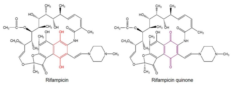
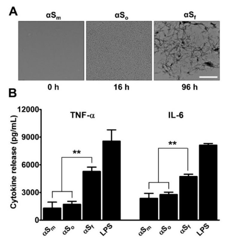
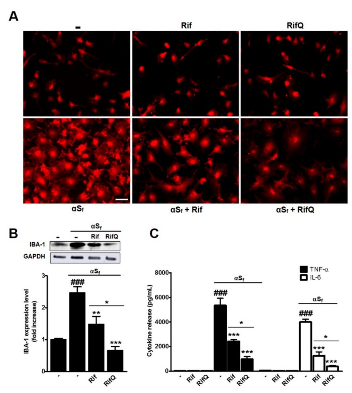
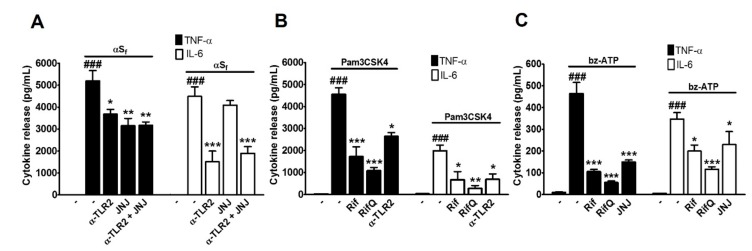
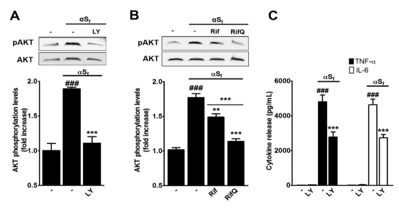
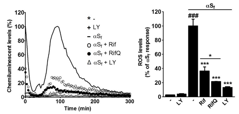
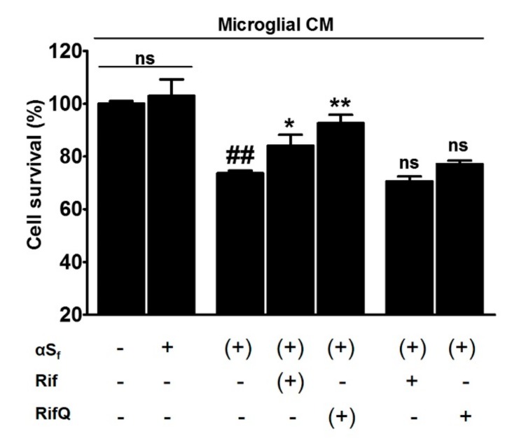
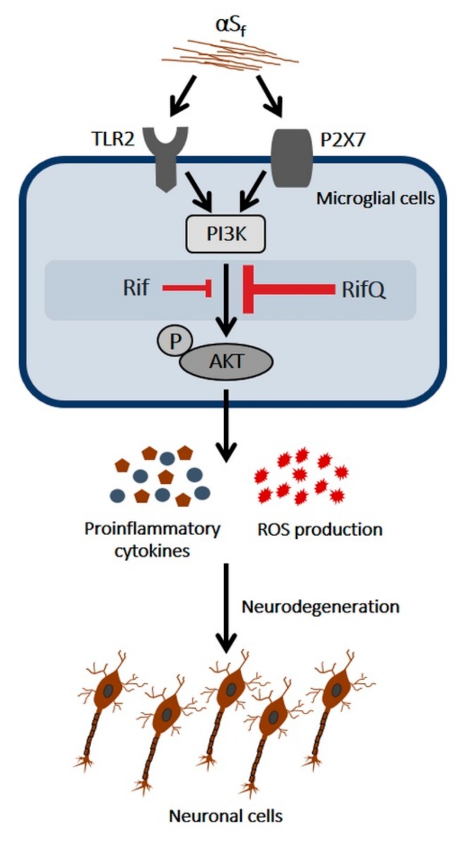
: Aggregated forms of the synaptic protein α-synuclein (αS) have been proposed to operate as a molecular trigger for microglial inflammatory processes and neurodegeneration in Parkinson´s disease. Here, we used brain microglial cell cultures activated by fibrillary forms of recombinant human αS to assess the anti-inflammatory and neuroprotective activities of the antibiotic rifampicin (Rif) and its autoxidation product rifampicin quinone (RifQ). Pretreatments with Rif and RifQ reduced the secretion of prototypical inflammatory cytokines (TNF-, IL-6) and the burst of oxidative stress in microglial cells activated with αS fibrillary aggregates. Note, however, that RifQ was constantly more efficacious than its parent compound in reducing microglial activation. We also established that the suppressive effects of Rif and RifQ on cytokine release was probably due to inhibition of both PI3K- and non-PI3K-dependent signaling events. The control of oxidative stress appeared, however, essentially dependent on PI3K inhibition. Of interest, we also showed that RifQ was more efficient than Rif in protecting neuronal cells from toxic factors secreted by microglia activated by αS fibrils. Overall, data with RifQ are promising enough to justify further studies to confirm the potential of this compound as an anti-parkinsionian drug.

Keywords: Parkinson’s disease; aggregation; cytokines; microglia; neuroinflammation; neuronal survival; α-synuclein.


Conflict of interest statement

The authors declare no conflict of interest.

Figures

Figure 1
Figure 1
Rif and RifQ chemical structures. The naphthyl core structure of Rif becomes a naphtoquinone in RifQ. This signifies that the para-diphenol (red) of the naphthyl core is converted into a para-quinone (violet).
Figure 2
Figure 2
Impact of α-synuclein species on microglial cytokine release. (A) Transmission electron microscopy images of αS (70 µg/ml) samples agitated at 37 °C by orbital shaking and harvested after 16 h or 96 h. Images show the presence of oligomers (αSo) and fibrillary species (αSf), at 16 and 96 h, respectively. Scale bar: 1 µm. (B) Impact that samples of αS (70 µg/mL) harvested after 16 h (αSo) or 96 h (αSf) of orbital agitation and non-shaken samples (αSm) have on the release of tumor necrosis factor (TNF)-α and Interleukin (IL)-6 in microglial cell cultures. Cytokine release was near to zero in control cultures. Lipopolysaccharide (LPS) (10 ng/mL) was utilized as a positive control for stimulation. Data are means ± standard error of the mean (SEM) of three independent experiments performed in triplicate. Note: **p < 0.01 vs. αSf.
Figure 3
Figure 3
Inhibitory effects of Rif and RifQ on αSf-activated microglial cells. (A) Visualization of the effects of Rif and RifQ (100 µM) on anti-ionized calcium binding adaptor molecule-1 (IBA-1) (red) immunofluorescent signals in microglial cells stimulated or not with αSf. A treatment with Rif or RifQ reduced the increase in IBA-1 expression observed in cultures exposed to αSf. Rif and RifQ had and no effect on basal IBA-1 expression levels. Scale bar: 20 µm. (B) Western blot analysis showing the extent of the inhibitory effects of Rif and RifQ on IBA-1 expression in microglial cells exposed to αSf. A representative blot is included (top), and relative intensity levels of IBA-1 are shown (bottom). (C) Effect of 100 µM Rif and RifQ on the amounts of TNF-α (filled bars) and IL-6 (open bars) released by microglial cells stimulated by αSf. Values are shown as means ± SEM of four independent experiments performed in triplicate. Note: ###p < 0.001 vs. controls, *p < 0.05 Rif vs. RifQ. **p < 0.01 and ***p < 0.001 vs. αSf.
Figure 4
Figure 4
Inhibitory effects of Rif and RifQ on microglial cells activated by either αSf or agonists of putative receptors for αSf. (A) Modulation of the effects of αSf by α-TLR2 (2.5 μg/ml), an antagonistic antibody against TLR2 and by JNJ 47965567 (JNJ) (20 μM), a synthetic antagonist of P2X7 receptors. (B) Modulation of the effects of Pam3CSK (1 µg/ml) on TNF-α and IL-6 release in microglial cell cultures treated with Rif, RifQ (both at 100 µM), or the TLR2 receptor antagonist (α-TLR2; 2.5 µg/ml). (C) Impact of a treatment with bz-ATP (500 µM), a specific agonist for P2X7 receptors, on TNF-α and IL-6 release in microglial cell cultures treated with Rif, RifQ (both at 100 µM), or the P2X7 receptor antagonist JNJ (20 µM). Values are shown as means ± SEM of four independent experiments performed in triplicate. Note: ###p < 0.001 vs. control, *p < 0.05, **p < 0.01, and ***p < 0.001 vs. corresponding inflammogen, i.e., αSf, Pam3CSK4, or bz-ATP.
Figure 5
Figure 5
Rif and RifQ inhibit a PI3K-dependent mechanism. (A) Western blot analysis of protein kinase B (AKT) phosphorylation in control and αSf-treated cultures exposed or not to the PI3K-specific inhibitor LY (2.5 μM). (B) Western blot analysis of AKT phosphorylation in control and αSf-activated microglial cells treated or not with 100 μM of Rif or RifQ. A representative blot is included (top), and relative levels of pAKT/AKT are shown (bottom). (C) TNF-α (filled bars) and IL-6 (open bars) released by microglial cells pretreated or not with LY prior to stimulation with αSf. Values are shown as means ± SEM of four independent experiments performed in triplicate. Note: ###p < 0.001 vs. controls. **p < 0.01 vs. αSf and ***p < 0.001 vs. αSf or Rif vs. RifQ.
Figure 6
Figure 6
Rif and RifQ prevent reactive oxygen species production in microglial cells activated by αSf. Left panel shows a representative kinetic profile of superoxide formation as a percentage of the maximal response elicited by αSf. The light intensity was monitored continuously for 300 min. Experimental values in the right panel represent the means of the area under the curve in percentage of the maximal response elicited by αSf. Data are expressed as the mean ± SEM of four independent experiments performed in triplicate. Note: ###p < 0.001 vs. controls, *p < 0.05 Rif vs. RifQ, ***p < 0.001 vs. αSf.
Figure 7
Figure 7
Rif and RifQ attenuate neuronal death caused by microglia activated by αSf. Cortical neurons were cultured for 48 h in the presence of microglial conditioned medium (CM) obtained as described in the text. (+): Indicates that the treatment was supplied initially to microglial cultures, and was thus provided indirectly to neuronal cultures through CM. The CCK-8 assay was used to evaluate neuronal viability. Data represent means of three different experiments performed in triplicate and error bars show SEM. Results are expressed as percentage of control values in cortical cultures receiving CM from control microglial cultures. Note: ##p < 0.01 vs. control CM; *p < 0.05, **p < 0.01 vs. αSf-treated CM; ns = not significantly different from αSf-treated CM.
Figure 8
Figure 8
Simplified scheme describing how Rif and RifQ attenuate the inflammatory response of microglial cells exposed to αSf. Microglial TLR2 and P2X7 receptors recognize fibrils of αS, which results in AKT phosphorylation by PI3K. This leads to stimulation of cytokine release and to a burst of oxidative stress, presumably by activation of the reduced nicotinamide adenine dinucleotide phosphate oxidase enzyme. Both pro-inflammatory cytokines and ROS are potentially deleterious for neuronal cells. The suppressive effect of Rif and RifQ on cytokine release is probably due to inhibition of both PI3K and non-PI3K-dependent signaling events. The control of oxidative stress appears, however, essentially dependent on PI3K inhibition. The inhibitory effects that Rif and RifQ exert on activated microglia may provide indirect protection to neuronal cells. RifQ appears constantly more effective than Rif in reducing inflammatory-type reactions and in providing neuronal rescue.

References

    1. Forno L.S. Neuropathology of Parkinson’s disease. J. Neuropathol. Exp. Neurol. 1996;55:259–272. doi: 10.1097/00005072-199603000-00001. - DOI - PubMed
    1. Goedert M., Spillantini M.G., Del Tredici K., Braak H. 100 years of Lewy pathology. Nat. Rev. Neurol. 2013;9:13–24. doi: 10.1038/nrneurol.2012.242. - DOI - PubMed
    1. Maries E., Dass B., Collier T.J., Kordower J.H., Steece-Collier K. The role of alpha-synuclein in Parkinson’s disease: Insights from animal models. Nat. Rev. Neurosci. 2003;4:727–738. doi: 10.1038/nrn1199. - DOI - PubMed
    1. Lee H.-J., Bae E.-J., Lee S.-J. Extracellular α-Snuclein-a novel and crucial factor in Lewy body diseases. Nat. Rev. Neurol. 2014;10:92–98. doi: 10.1038/nrneurol.2013.275. - DOI - PubMed
    1. Glass C.K., Saijo K., Winner B., Marchetto M.C., Gage F.H. Mechanisms underlying inflammation in neurodegeneration. Cell. 2010;140:918–934. doi: 10.1016/j.cell.2010.02.016. - DOI - PMC - PubMed

Publication types

MeSH terms