. 2019 Jul 26;52(1):38.
doi: 10.1186/s40659-019-0245-4.

miR-29b-3p promotes progression of MDA-MB-231 triple-negative breast cancer cells through downregulating TRAF3

Affiliations

miR-29b-3p promotes progression of MDA-MB-231 triple-negative breast cancer cells through downregulating TRAF3

Bao Zhang et al. Biol Res. .

Abstract

Background: Breast cancer is the second common malignant cancer among females worldwide. Accumulating studies have indicated that deregulation of miRNA expression in breast cancer will contribute to tumorigenesis and form different cancer subtypes. However, the reported studies on miR-29b-3p-regulated breast cancer are limited so far. Herein, we investigated the role and mechanism of miR-29b-3p in the triple negative breast cancer cell line MDA-MB-231.

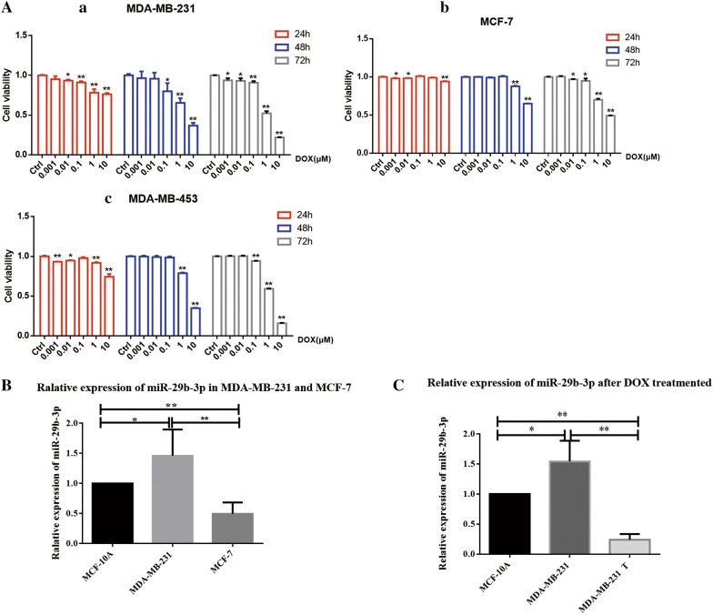
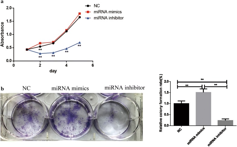
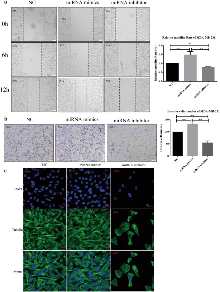
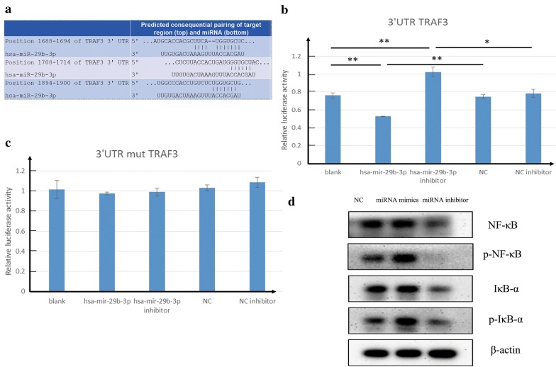
Methods: The relative miR-29b-3p expression in different breast cancer cell lines were determined by qRT-PCR. CCK8 and colony formation assay were used to determine the influence of miR-29b-3p on cell proliferation. Migration assay and invasion assay were performed for cell migration and invasion respectively. To study the cell integrity immunofluorescence was performed. TUNEL assay, flow cytometry assay, hoechst staining and western blot were conducted to determine the influence of miR-29b-3p inhibitor on cell apoptosis. TRAF3 was found to be the target gene of miR-29b-3p using bioinformatics predictions. Dual-luciferase assay was performed to determine the relative luciferase activity in NC, miR-29b-3p mimic, miR-29b-3p inhibitor with TRAF3 3'-UTR wt or TRAF3 3'-UTR mt reporter plasmids. The proteins expression of NF-κB signaling pathway in MDA-MB-231 after transfection with NC, miR-29b-3p mimic, miR-29b-3p inhibitor were determined by western blot.

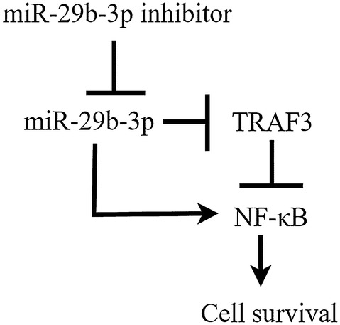
Results: The miR-29b-3p expression was significantly increased in MDA-MB-231 compare with MCF-10A. miR-29b-3p inhibitor reduced the cell viability of MDA-MB-231 and inhibited cell migration and invasion. Cell cytoskeleton integrity destroyed after miR-29b-3p inhibitor treatment. Furthermore, we identified the mechanism and found miR-29b-3p targets the TRAF3 and activates NF-κB signaling pathway.

Conclusions: From the above studies, our results indicated that miR-29b-3p acts as a promoter for the development of MDA-MB-231.

Keywords: Cytoskeleton; NF-κB; TRAF3; Triple negative breast cancer; miR-29b-3p.


Conflict of interest statement

The authors declare that they have no competing interests.

Figures

Fig. 1
Fig. 1
The expression of miR-29b-3p in breast cancer cells. A Doxorubicin exhibits cytotoxic effect in breast cancer cells. (a) MDA-MB-231, (b) MCF-7 and (c) MDA-MB-453. The percentage inhibition of cell viability represented graphically. B Analysis of miR-29b-3p expression level in MDA-MB-231 and MCF-7 cells compared with MCF-10A cells. C Analysis of miR-29b-3p expression level in MDA-MB-231 after Doxorubicin treated. U6 was used as an internal control. T treated. Data are presented as the mean ± SD of three independent experiments. *p < 0.05, **p < 0.01
Fig. 2
Fig. 2
Cell proliferation and colony forming ability of MDA-MB-231 cells. a CCK-8 assay was applied to determine the activity of cells after transfection. b miR-29b-3p inhibitor inhibited cell colony formation in MDA-MB-231 cells. Data are presented as the mean ± SD of three independent experiments. *p < 0.05, **p < 0.01
Fig. 3
Fig. 3
Cell migration and invasion of MDA-MB-231 cells. a miR-29b-3p inhibitor inhibited cell migration in the MDA-MB-231 cell lines. Image magnification: ×20. b miR-29b-3p inhibitor inhibited cell invasion in the MDA-MB-231 cell lines. Image magnification: ×20. c Immunofluorescence for cytoskeleton. The nuclei were visualized by staining with DAPI (blue). α-Tubulin (green). Image magnification: ×60. Data are presented as the mean ± SD of three independent experiments. *p < 0.05, **p < 0.01
Fig. 4
Fig. 4
miR-29b-3p inhibitor induces apoptosis. A TUNEL assay. The nuclei were visualized by staining with DAPI (blue), Cy3-labeled the apoptosis cells (red). Image magnification: ×20. B Annexin V-FITC and propidium iodide assay. The sum of early and late apoptotic cells ratio (%) were quantitated by flow cytometer analysis of Annexin V/PI. C Hoechst staining. (a) MDA-MB-231; (b) MCF-7. White arrows indicate the cells nuclear fragmentation. The nuclei were visualized by staining with Hoechst (red). Image magnification: ×40. D Western blot for Cleaved PARP. Data are presented as the mean ± SD of three independent experiments. The data represents three experiments exhibiting similar results
Fig. 5
Fig. 5
miR-29b-3p targets TRAF3 and Effects on NF-κB signaling pathway. a Alignment of the miR-29b-3p seed sequence with the TRAF3 3′ UTR. b TRAF3 3′-UTR wt relative luciferase activity. c TRAF3 3′-UTR mutant relative luciferase activity. d Western blot for NF-κB pathway. β-Actin was used as used as an internal control. Data are presented as the mean ± SD of three independent experiments. *p < 0.05, **p < 0.01
Fig. 6
Fig. 6
Schematic representation of miR-29b-3p inhibitor regulated miR-29b-3p mediated NF-κB pathway

References

    1. Banerji S, et al. Sequence analysis of mutations and translocations across breast cancer subtypes. Nature. 2012;486(7403):405–409. - PMC - PubMed
    1. Siegel RL, Miller KD, Jemal A. Cancer statistics, 2017. CA Cancer J Clin. 2017;67:7–30. - PubMed
    1. Fertig EJ, et al. Analysis of gene expression of secreted factors associated with breast cancer metastases in breast cancer subtypes. Sci Rep. 2015;5:12133. - PMC - PubMed
    1. Fan L, et al. Breast cancer in China. Lancet Oncol. 2014;15(7):e279–e289. - PubMed
    1. Survcan WHO. Cancer survival in Qidong C, 1992–2000. http://survcan.iarc.fr/survival/chap7.pdf. Accessed 15 Mar 2019.

MeSH terms