. 2019 Jul 26;7(1):120.
doi: 10.1186/s40478-019-0777-4.

Soluble aggregates present in cerebrospinal fluid change in size and mechanism of toxicity during Alzheimer's disease progression

Affiliations

Soluble aggregates present in cerebrospinal fluid change in size and mechanism of toxicity during Alzheimer's disease progression

Suman De et al. Acta Neuropathol Commun. .

Abstract

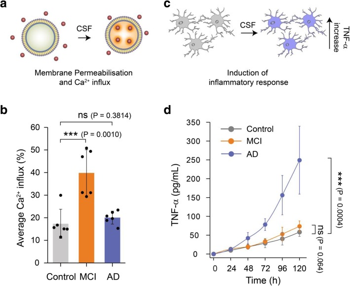
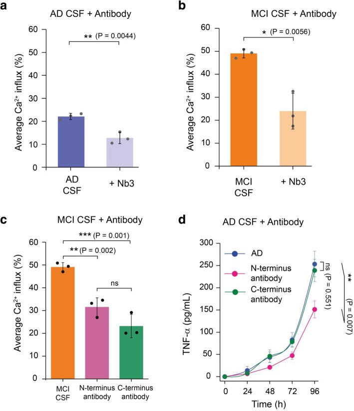
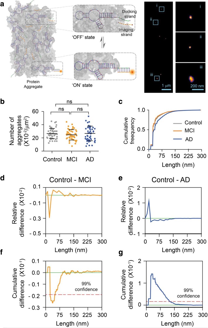
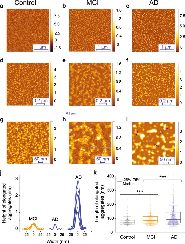
Soluble aggregates of amyloid-β (Aβ) have been associated with neuronal and synaptic loss in Alzheimer's disease (AD). However, despite significant recent progress, the mechanisms by which these aggregated species contribute to disease progression are not fully determined. As the analysis of human cerebrospinal fluid (CSF) provides an accessible window into the molecular changes associated with the disease progression, we characterised soluble aggregates present in CSF samples from individuals with AD, mild cognitive impairment (MCI) and healthy controls using a range of sensitive biophysical methods. We used super-resolution imaging and atomic force microscopy to characterise the size and structure of the aggregates present in CSF and correlate this with their ability to permeabilise lipid membranes and induce an inflammatory response. We found that these aggregates are extremely heterogeneous and exist in a range of sizes, varying both structurally and in their mechanisms of toxicity during the disease progression. A higher proportion of small aggregates of Aβ that can cause membrane permeabilization are found in MCI CSF; in established AD, a higher proportion of the aggregates were larger and more prone to elicit a pro-inflammatory response in glial cells, while there was no detectable change in aggregate concentration. These results show that large aggregates, some longer than 100 nm, are present in the CSF of AD patients and suggest that different neurotoxic mechanisms are prevalent at different stages of AD.

Keywords: Alzheimer’s disease; Cerebrospinal fluid; Disease mechanism; Mild cognitive impairment; Protein aggregation; Structure-function relation; Super-resolution imaging.


Conflict of interest statement

CEB is a member of the GSK Immunology Catalyst, serves on the scientific advisory board of Nodthera, is a consultant for Syncona and a co-founder of Polypharmakos. KB has served as a consultant or at advisory boards for Alector, Alzheon, CogRx, Biogen, Lilly, Novartis and Roche Diagnostics, and is a co-founder of Brain Biomarker Solutions in Gothenburg AB, a GU Ventures-based platform company at the University of Gothenburg, IS has served as a consultant for Takeda. HZ has served at scientific advisory boards for Roche Diagnostics, Wave, Samumed and CogRx and is a co-founder of Brain Biomarker Solutions in Gothenburg AB, a GU Ventures-based platform company at the University of Gothenburg, all unrelated to the work presented in this paper. All the other authors declare no competing interests.

Figures

Fig. 1
Fig. 1
Soluble aggregates present in MCI and AD CSF samples show different dominant mechanisms of toxicity. a Membrane permeabilisation was measured by immobilising hundreds of vesicles containing a Ca2+-sensitive dye on PEGylated glass cover slides. If any species present in the CSF disrupts the integrity of the lipid membrane of the vesicles, Ca2+ ions from the surrounding buffer enter into individual vesicles in numbers that can be quantified using highly sensitive TIRF microscopy. b Aliquots of MCI CSF can cause more membrane permeabilisation compared to AD and control CSF (n = 6 AD, 6 MCI, 6 control CSF). A two-sample unpaired t-test was performed to compare each data set. c The inflammatory response in microglia cells was quantified using an ELISA assay to measure the levels of secreted tumour necrosis factor alpha (TNF-ɑ). d For this study, CSF samples were added to BV2 microglia cells and incubated for 120 h. Every 24 h the TNF-α concentration in the supernatant was quantified using an ELISA assay. AD CSF samples were more effective MCI and control CSF samples in inducing an inflammatory response (lines are guide to the eye; n = 10 AD, 6 MCI, 6 control CSF). Error bars are the standard deviation among data points. A two-sample unpaired t-test at the 120 h time point was performed to compare the data sets
Fig. 2
Fig. 2
The toxic soluble aggregates present in MCI and AD CSF contain Aβ. A significant inhibition of the membrane permeabilisation of lipid membranes by (a) AD and (b) MCI CSF samples is caused by a nanobody Nb3 (300 nM) designed to bind to Aβ, indicating that some of the aggregates present in MCI and AD CSF contain Aβ. Nb3 recognises amino acids 17–28 of the Aβ sequence and shown to inhibit toxicity induced by protein aggregates of Aβ cerebrospinal fluid samples from AD patient (c) Both N-terminally and C-terminally targeting antibodies significantly inhibit MCI CSF-induced membrane permeabilisation. However, we do not find any significant difference between their activities. Error bars are the standard deviation among data points. Two sample unpaired t-tests were performed to compare the data sets (n = 3). d A N-terminal binding antibody (binding the region of residues 3–9 of Aβ42) is more potent at reducing the aggregate-induced inflammatory response than a C-terminal designed antibody (binding the region of residues 36–42 of Aβ42); P values are calculated using a two sample t-test to compare the inhibition by an N-terminally binding antibody and a C-terminally antibody at 96 h (n = 3). Error bars are the standard deviation among data points
Fig. 3
Fig. 3
Super-resolution imaging of aggregates present in CSF using AD-PAINT. a Schematic of AD PAINT. Dye-labelled DNA imaging strands transiently bind to their complementary target sequence (docking strand), which is attached to a protein aggregate via aptamer. This transient binding between imaging and docking strand is detected. Repeated cycles of binding and unbinding allows a super-resolved image of individual protein aggregate present in CSF to be determined. The right image shows examples of super-resolved image of protein aggregates from CSF. Three individual protein aggregates present in CSF are enlarged. The lengths of the aggregates shown are: (i) 47 nm, (ii) 34 nm and (iii) 118 nm. b Number of aggregates present in control, MCI and AD CSF samples. Each point represents one field of view. c Cumulative frequency histograms of the size distributions of all aggregates measured. d, e Differences between normalised histograms of the size distributions of the indicated CSF samples. f, g Differences between the cumulative frequency histograms of the size distributions of the indicated CSF samples. The dotted line indicates 99% confidence using the Kolmogorov-Smirnov statistical test. (n = 6 AD, 6 MCI, 6 control CSF)
Fig. 4
Fig. 4
Characterization of the protein aggregates present in CSF samples at single aggregate level using AFM. a-c Representative AFM images of the aggregates present at different CSF samples with (d-j) a magnified example of the species present. j, k Statistical analysis of the cross-sectional length and heights of individual aggregates present in CSF samples; the length of the individual aggregates are shown as a box chart

References

    1. Aprile Francesco A., Sormanni Pietro, Perni Michele, Arosio Paolo, Linse Sara, Knowles Tuomas P. J., Dobson Christopher M., Vendruscolo Michele. Selective targeting of primary and secondary nucleation pathways in Aβ42 aggregation using a rational antibody scanning method. Science Advances. 2017;3(6):e1700488. doi: 10.1126/sciadv.1700488. - DOI - PMC - PubMed
    1. Benilova I, Karran E, De Strooper B. The toxic Aβ oligomer and Alzheimer’s disease: an emperor in need of clothes. Nat Neurosci. 2012;15:349–357. doi: 10.1038/nn.3028. - DOI - PubMed
    1. Bjerke M, Kern S, Blennow K, Zetterberg H, Waern M, Börjesson-Hanson A, et al. Cerebrospinal Fluid Fatty Acid-Binding Protein 3 is Related to Dementia Development in a Population-Based Sample of Older Adult Women Followed for 8 Years. J Alzheimer’s Dis. 2016;49:733–741. doi: 10.3233/JAD-150525. - DOI - PubMed
    1. Blennow K. Cerebrospinal fluid protein biomarkers for Alzheimer’s disease. NeuroRX. 2004;1:213–225. doi: 10.1602/neurorx.1.2.213. - DOI - PMC - PubMed
    1. Blennow K, Hampel H, Weiner M, Zetterberg H. Cerebrospinal fluid and plasma biomarkers in Alzheimer disease. Nat rev Neurol. 2010;6:131. doi: 10.1038/nrneurol.2010.4. - DOI - PubMed

Publication types