Review
. 2019 Sep:37:100590.
doi: 10.1016/j.blre.2019.100590. Epub 2019 Jul 16.

Update on mutations in the HIF: EPO pathway and their role in erythrocytosis

Affiliations
Review

Update on mutations in the HIF: EPO pathway and their role in erythrocytosis

Terence R Lappin et al. Blood Rev. 2019 Sep.

Abstract

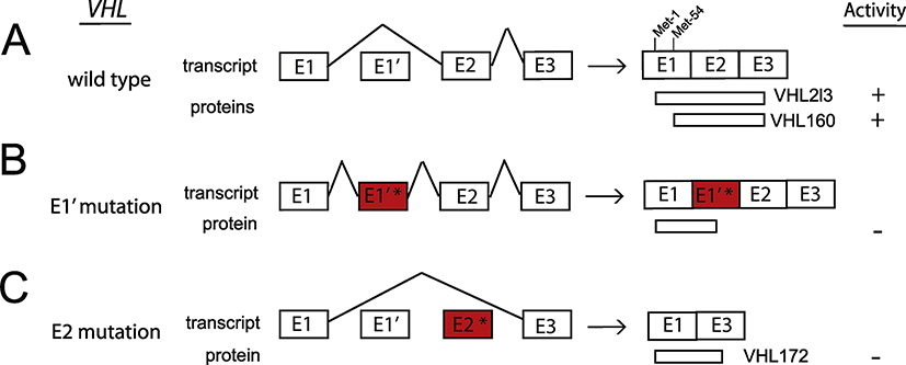
Identification of the underlying defects in congenital erythrocytosis has provided mechanistic insights into the regulation of erythropoiesis and oxygen homeostasis. The Hypoxia Inducible Factor (HIF) pathway plays a key role in this regard. In this pathway, an enzyme, Prolyl Hydroxylase Domain protein 2 (PHD2), constitutively prolyl hydroxylates HIF-2α, thereby targeting HIF-2α for degradation by the von Hippel Lindau (VHL) tumor suppressor protein. Under hypoxia, this modification is attenuated, resulting in the stabilization of HIF-2α and transcriptional activation of the erythropoietin (EPO) gene. Circulating EPO then binds to the EPO receptor (EPOR) on red cell progenitors in the bone marrow, leading to expansion of red cell mass. Loss of function mutations in PHD2 and VHL, as well as gain of function mutations in HIF-2α and EPOR, are well established causes of erythrocytosis. Here, we highlight recent developments that show that the study of this condition is still evolving. Specifically, novel mutations have been identified that either change amino acids in the zinc finger domain of PHD2 or alter splicing of the VHL gene. In addition, continued study of HIF-2α mutations has revealed a distinctive genotype-phenotype correlation. Finally, novel mutations have recently been identified in the EPO gene itself. Thus, the cascade of genes that at a molecular level leads to EPO action, namely PHD2 - > HIF2A - > VHL - > EPO - > EPOR, are all mutational targets in congenital erythrocytosis.

Keywords: Erythrocytosis; Erythropoiesis; Erythropoietin; Hypoxia Inducible Factor; Oxygen sensing; Polycythemia; Prolyl Hydroxylase Domain protein; Zinc finger; von Hippel Lindau tumor suppressor protein.


Conflict of interest statement

Conflict of interest

No conflicts to disclose.

Figures

Fig. 1.
Fig. 1.. The feedback loop that controls red blood cell production.
The oxygen sensing mechanism which regulates EPO production in the kidney is illustrated in the top left panel. In normoxic conditions, PHD2 hydroxylates one or both of a pair of highly conserved prolines in HIF-2α in an oxygen-dependent manner, using 2-oxoglutarate and ascorbate as co-factors. This hydroxylation enables specific binding by a ubiquitin ligase complex containing VHL, elongin B (B) and elongin C (C) which leads to ubiquitination and subsequent proteasomal degradation (for simplicity, two other components of the complex, cullin-2 and rbx1, are omitted). Under hypoxia the hydroxylation of HIF-2α is attenuated, permitting it to bind to the HRE in 5’ region of the EPO gene thereby enabling enhanced EPO transcription. EPO enters the circulation and binds to EPO receptors on erythroid progenitor cells (primarily Colony Forming Unit-Erythroid cells) inducing the proliferation and differentiation of erythroid cells (top right panel). The circulating red blood cell (RBC) mass is therefore dependent on the degree of hypoxia sensed by PHD2 in the kidney, and reflects the dynamic balance between EPO-induced red cell production and subsequent loss or destruction of mature red blood cells.
Fig. 2.
Fig. 2.. The PHD2-HIF-VHL-EPO axis and its dysregulation.
(A) Scheme depicting the distinctive oxygen-sensing mechanism that regulates EPO transcription. In the presence of oxygen, PHD2 site-specifically hydroxylates HIF-2α, thereby targeting it for degradation by VHL. Little HIF-2α remains to bind the HRE of the EPO gene, so activation is modest. ARNT is the stable subunit of HIF-2. (B) Loss-of-function mutations in PHD2, denoted by PHD2*, cause an increase in HIF-2α and increased (or inappropriately normal) EPO gene activation, designated erythrocytosis type 3 (ECYT3) in the OMIM classification. (C) In gain-of-function mutations in HIF-2α, denoted by HIF-2α*, hydroxylation is deceased and VHL binding is reduced, leading to increased EPO gene activation, ECYT4. (D) Loss-of-function mutations in VHL, denoted by VHL*, reduce binding to hydroxylated HIF-2α leading to increased EPO gene activation, ECYT2. (E) A mutation in EPO, denoted by EPO* causes a frameshift that initiates excess production of EPO from a normally noncoding EPO mRNA, ECYT5.
Fig. 3.
Fig. 3.. Loss of function mutations in PHD2 cause ECYT3.
(A) Diagram of PHD2 depicting the location of the zinc finger and prolyl hydroxylase domains. The sequence of the zinc finger domain across various metazoan species is shown with the areas shaded in gray denoting the zinc-binding residues. The recently reported mutations Y41C and C42R mutations cause erythrocytosis [44]. Earlier erythrocytosis-associated mutations in PHD2 reside within or near the prolyl hydroxylase domain and comprise missense (circles), nonsense (squares), and frameshift (triangles) mutations. Numbers at top and bottom indicate residue number. (B) Model showing recruitment of PHD2 to the HSP90 complex by interaction of the PHD2 zinc finger (ZF) with a PXLE motif in p23, an HSP90 cochaperone that interacts directly with HSP90. This facilitates hydroxylation of HIF-α, a client protein of the HSP90 complex.
Fig. 4.
Fig. 4.. Gain of function mutations in HIF-2α cause ECYT4 and neuroendocrine tumors.
Diagram of HIF-2α depicting the location of the oxygen dependent degradation domain (ODDD), as well as the basic helix-loop-helix (bHLH), Per-Arnt-Sim A (PAS-A), PAS-B, and C-terminal activation domain (CAD) domains. Patients who presented with PPGL combined with somatostatinoma and erythrocytosis, PPGL with erythrocytosis, PPGL only, and erythrocytosis only are shown together with the frequency of these disorders found caused by each mutation reported [80] denoted by colored circles. All HIF-2α mutations reported so far in the context of erythrocytosis are heterozygous and the majority are missense mutations. The underlined red “P” indicates Pro-531, the primary site of hydroxylation in HIF-2α. Numbers at bottom indicate residue number.
Fig. 5.
Fig. 5.. Loss of function mutations in VHL cause ECYT2 and can be due to aberrant splicing.
(A) Wild type VHL contains 3 canonical exons (E1, E2, E3) that encode the functionally active 213 aa protein, VHL213 (also known as p30). VHL E1 contains an internal translation initiation codon, Met-54, that causes the production of a truncated form, VHL160 (also known as p19). Lenglet and colleagues (2018) reported a new spliced isoform that contains a cryptic exon E1’. Exons are not drawn to scale. (B) Mutations in E1’ (denoted by E1’*) result in inclusion of E1’. The isoform containing E1 spliced with E1’ may theoretically encode a protein of 193 aa, of which 114 aa are encoded by E1, and 79 aa by E1’ and is functionally inactive. (C) Mutations in E2 (denoted by E2*) result in splicing of E1 to E3. The isoform containing E1 spliced with E3 encodes VHL172 which lacks E2 and is inactive.
Fig. 6.
Fig. 6.. Gain of function mutation in EPO cause ECYT5 and are due to a single nucleotide deletion in Exon 2.
(A) Wild type EPO mRNA, transcribed from promoter P1, contains 5 exons (E1-E5) that encode a protein of 194 aa consisting of a 27 aa signal peptide and the functionally active 167 aa mature protein. The sequence of the first 14 amino acids of the signal peptide is shown. An alternative promoter located in intron 1, P2, produces a transcript that contains 4 exons which does not make functional EPO. Single underlined green AUG = AUG1, double underlined blue AUG = AUG2. Exons are not drawn to scale. (B) Zmajkovic et al (2018) [97] found that a single-nucleotide deletion introduces a frameshift in E2 (denoted by E2*) that interrupts translation of the normal EPO mRNA transcribed from P1 (producing a short non-functional peptide terminating in exon 3), but instead initiates excess functionally active EPO from the normally noncoding transcript from the alternative P2 promoter utilizing AUG2. The location of the G that is deleted in Exon 2 is shown in red and by triangle in panel A and the sequence of the first 9 amino acids is shown. In essence, the P2 transcripts produce polypeptides arising from the alternative translation initiation site in Exon 2 that are now in the proper reading frame with respect to the remainder of the EPO coding sequence and are responsible for the overproduction of EPO leading to erythrocytosis. The signal peptide encoded by the E2 mutant is 22 aa, 5 aa shorter than wild type EPO signal peptide.

References

    1. Wenger RH, Hoogewijs D. Regulated oxygen sensing by protein hydroxylation in renal erythropoietin-producing cells. Am J Physiol Renal Physiol. 2010;298:F1287–96. - PubMed
    1. Haase VH. Regulation of erythropoiesis by hypoxia-inducible factors. Blood Rev. 2013;27:41–53. - PMC - PubMed
    1. Suzuki N, Yamamoto M. Roles of renal erythropoietin-producing (REP) cells in the maintenance of systemic oxygen homeostasis. Pflugers Arch. 2016;468:3–12. - PubMed
    1. Kaelin WG Jr., Ratcliffe PJ. Oxygen sensing by metazoans: the central role of the HIF hydroxylase pathway. Mol Cell. 2008;30:393–402. - PubMed
    1. Majmundar AJ, Wong WJ, Simon MC. Hypoxia-inducible factors and the response to hypoxic stress. Mol Cell. 2010;40:294–309. - PMC - PubMed

Publication types