. 2020 May 1;116(6):1199-1213.
doi: 10.1093/cvr/cvz199.

Exercise restores dysregulated gene expression in a mouse model of arrhythmogenic cardiomyopathy

Affiliations

Exercise restores dysregulated gene expression in a mouse model of arrhythmogenic cardiomyopathy

Sirisha M Cheedipudi et al. Cardiovasc Res. .

Abstract

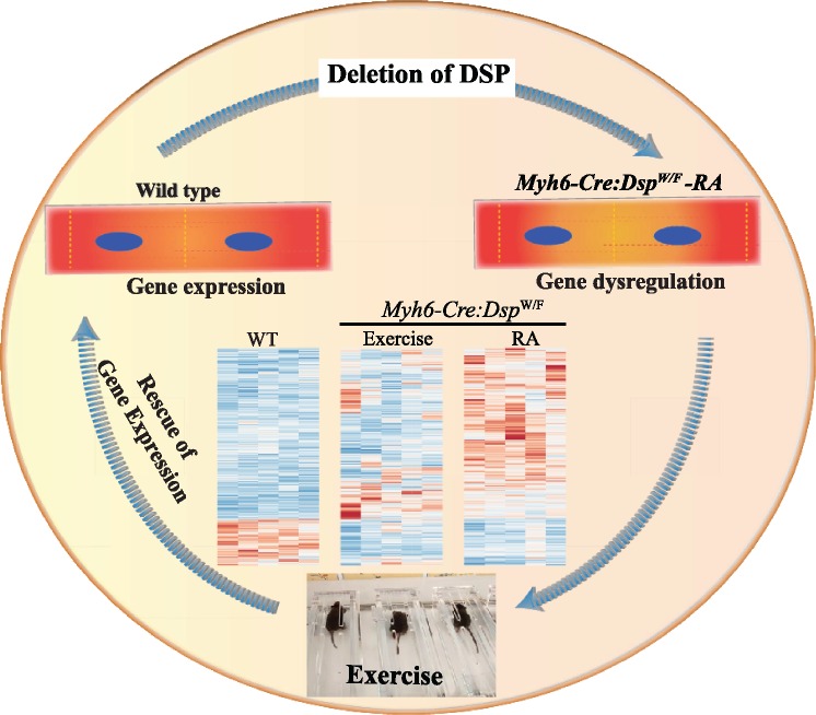
Aims: Arrhythmogenic cardiomyopathy (ACM) is a myocardial disease caused mainly by mutations in genes encoding desmosome proteins ACM patients present with ventricular arrhythmias, cardiac dysfunction, sudden cardiac death, and a subset with fibro-fatty infiltration of the right ventricle predominantly. Endurance exercise is thought to exacerbate cardiac dysfunction and arrhythmias in ACM. The objective was to determine the effects of treadmill exercise on cardiac phenotype, including myocyte gene expression in myocyte-specific desmoplakin (Dsp) haplo-insufficient (Myh6-Cre:DspW/F) mice.

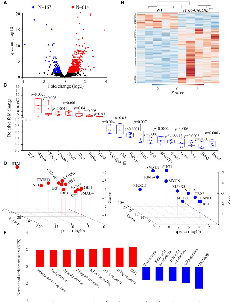
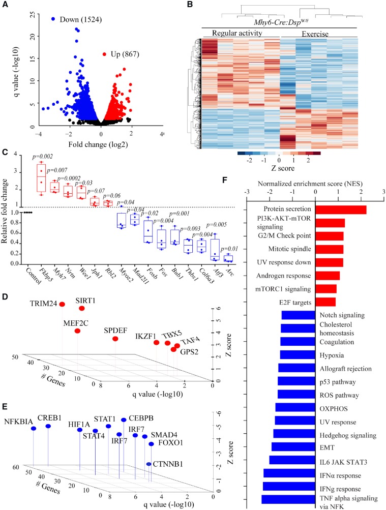
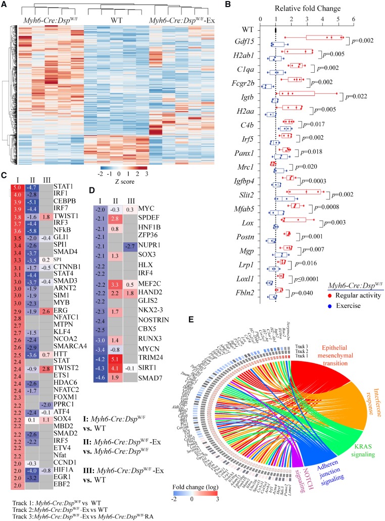
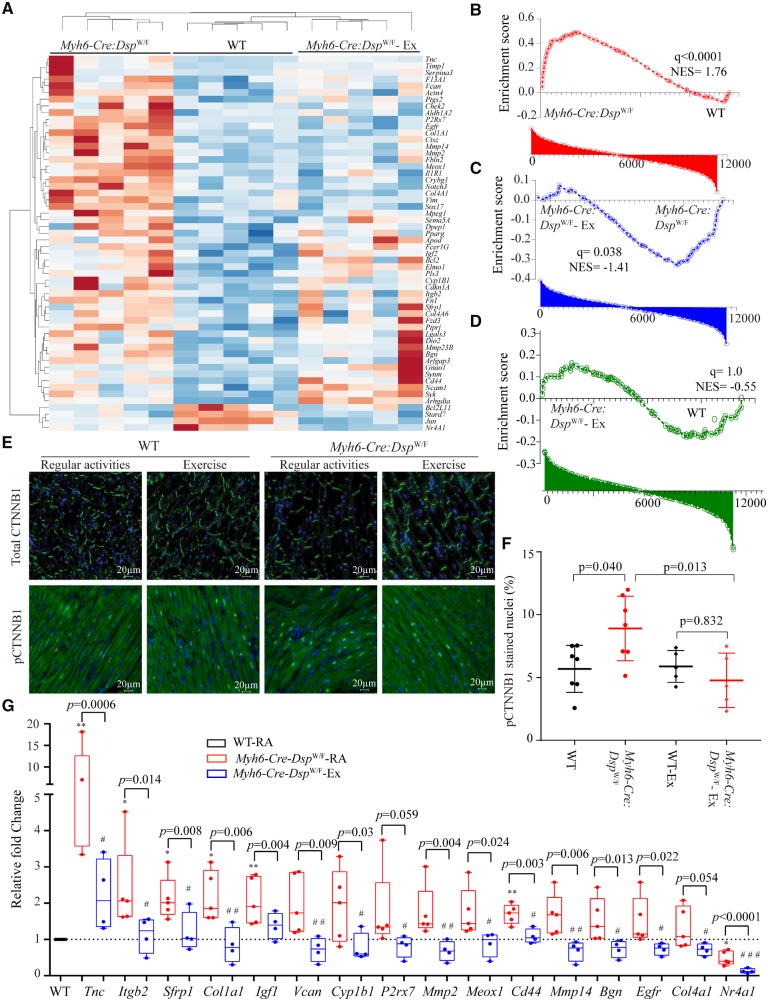
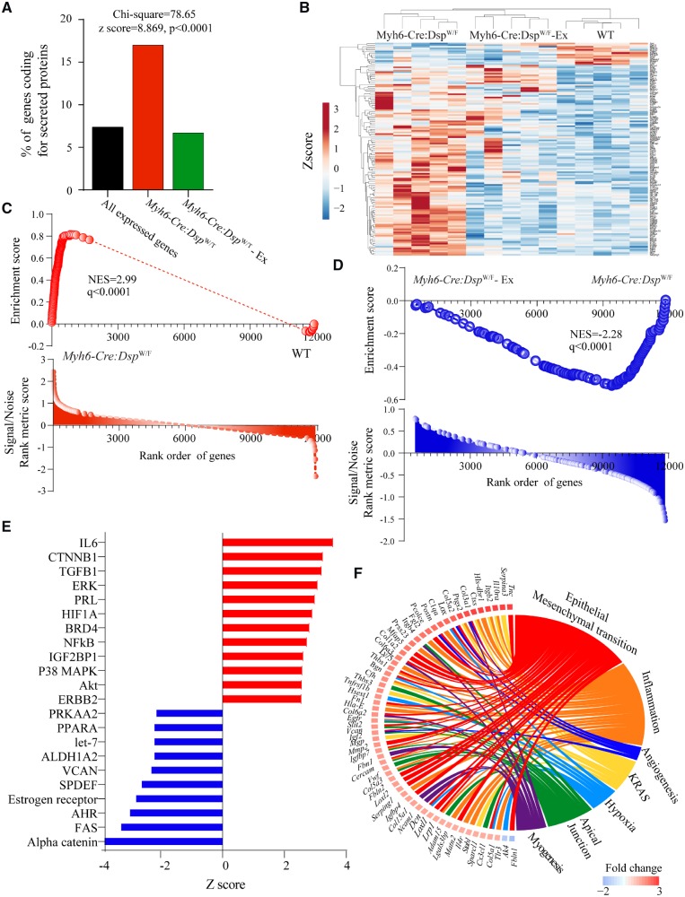
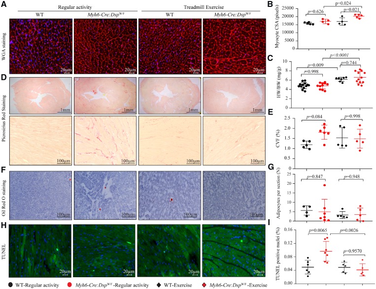
Methods and results: Three months old sex-matched wild-type (WT) and Myh6-Cre:DspW/F mice with normal cardiac function, as assessed by echocardiography, were randomized to regular activity or 60 min of daily treadmill exercise (5.5 kJ work per run). Cardiac myocyte gene expression, cardiac function, arrhythmias, and myocardial histology, including apoptosis, were analysed prior to and after 3 months of routine activity or treadmill exercise. Fifty-seven and 781 genes were differentially expressed in 3- and 6-month-old Myh6-Cre:DspW/F cardiac myocytes, compared to the corresponding WT myocytes, respectively. Genes encoding secreted proteins (secretome), including inhibitors of the canonical WNT pathway, were among the most up-regulated genes. The differentially expressed genes (DEGs) predicted activation of epithelial-mesenchymal transition (EMT) and inflammation, and suppression of oxidative phosphorylation pathways in the Myh6-Cre:DspW/F myocytes. Treadmill exercise restored transcript levels of two-third (492/781) of the DEGs and the corresponding dysregulated transcriptional and biological pathways, including EMT, inflammation, and secreted inhibitors of the canonical WNT. The changes were associated with reduced myocardial apoptosis and eccentric cardiac hypertrophy without changes in cardiac function.

Conclusion: Treadmill exercise restored transcript levels of the majority of dysregulated genes in cardiac myocytes, reduced myocardial apoptosis, and induced eccentric cardiac hypertrophy without affecting cardiac dysfunction in a mouse model of ACM. The findings suggest that treadmill exercise has potential beneficial effects in a subset of cardiac phenotypes in ACM.

Keywords: Arrhythmogenic cardiomyopathy; Exercise; Gene expression.


Figures

Graphical Abstract
Graphical Abstract
Figure 1
Figure 1
Cardiac myocyte transcriptome in Myh6-Cre:DspW/F mice. (A) Volcano plot showing down-regulated and up-regulated genes in cardiac myocytes isolated from 6 months old Myh6-Cre:DspW/F as compared to wild type (WT) mice (N = 5 per group). (B) Heat map of the differentially expressed genes (DEGs). (C) Quantitative PCR validation of the top up-regulated and down-regulated genes in cardiac myocytes isolated from 6-month-old wild type and Myh6-Cre:DspW/F mice (N = 5). (D and E) Transcriptional factors (TFs) predicted to be activated (C) and suppressed (D) in Myh6-Cre:DspW/F myocytes. The X-axis represents −log10 q value, the Y-axis represents Z score, and the Z-axis represents number of target genes of the transcription factor present in the differentially expressed genes with q < 0.05. (F) Dysregulated biological pathways corresponding to the DEGs with q < 0.05 (Y-axis represents Z score). P-values were calculated by t-test for normally distributed data and Mann–Whitney test for those departing from normality.
Figure 2
Figure 2
Effects of treadmill exercise on cardiac myocyte transcriptome. (A) Volcano plot showing down-regulated and up-regulated genes in cardiac myocytes isolated from the hearts of Myh6-Cre:DspW/F mice in the treadmill exercise group as opposed to the regular activity group (N = 5 per group). (B) Heat map of DEGs between indicated groups showing clustering based on the genotypes. (C) Quantitative PCR data for top dysregulated genes in isolated cardiac myocytes from Myh6-Cre:DspW/F in the regular activity group (N = 5) and Myh6-Cre:DspW/F in the treadmill exercise group (N = 4). (D and E) TFs predicted to be activated (D) and suppressed (E) based on DEGs with a q < 0.05 in Myh6-Cre:DspW/F myocytes in the treadmill exercise group as compared to the regular activity group. The X-axis represents −log10 of q value, the Y-axis represents Z score, and the Z-axis represents number of genes predicted to be targets of each TF. (F) Dysregulated biological pathways corresponding to DEGs with q < 0.05. Pathways with q < 0.05 and normalized enrichment score, in the X-axis, are shown. P-values were calculated by t-test for normally distributed data and Mann–Whitney test for those departing from normality.
Figure 3
Figure 3
Rescue of cardiac myocyte transcriptome, TFs and biological pathways in Myh6-Cre:DspW/F mice in the exercise group. (A) Heat map of DEGs in Myh6-Cre:DspW/F mice assigned to the regular activity, WT mice, and Myh6-Cre:DspW/F mice assigned to treadmill exercise are shown. The map shows clustering of the transcripts in Myh6-Cre:DspW/F myocytes in the exercise group with those in the WT myocytes. (B) Transcript levels of selected DEGs corresponding to panel A, as determined by qPCR (one way ANOVA P-values are shown). The panel shows normalization of the transcript levels, similar to levels in the WT, in the Myh6-Cre:DspW/F in the treadmill exercise group. (C and D) Heat maps and Z scores showing rescue and normalization of all activated (C) and suppressed (D) TFs in Myh6-Cre:DspW/F myocytes upon treadmill exercise. Pairwise comparisons are shown; I: Myh6-Cre:DspW/F regular activity (RA) vs. WT; II: Myh6-Cre:DspW/F—treadmill exercise (Ex) vs. Myh6-Cre:DspW/F—RA; III: Myh6-Cre:DspW/F—Ex vs. WT. (E) Circos map showing rescue of DEGs and their corresponding enriched Hallmark pathways.
Figure 4
Figure 4
Rescue of the canonical WNT pathway in Myh6-Cre:DspW/F mice upon treadmill exercise. (A) Heat map of selected known canonical WNT target gene transcripts in Myh6-Cre:DspW/F myocytes and their clustering with the transcripts in WT myocytes with exercise. (BD) Pairwise gene set enrichment analysis plots and the corresponding q values, showing enrichment of transcript levels of the canonical WNT pathway target genes and the effects of exercise. (B) Myh6-Cre:DspW/F vs. WT myocytes; (C) Myh6-Cre:DspW/F—regular activity vs. Myh6-Cre:DspW/F—exercise; (D) Myh6-Cre:DspW/F—exercise vs. WT myocytes. (E) Representative immunofluorescence panels showing expression and subcellular localization of total and phospho-β catenin (Ser33/37/Thr41) in the experimental groups (N = 7 in WT and Myh6-Cre:DspW/F regular activity groups and N = 5 mice for WT and Myh6-Cre:DspW/F - exercise groups). (F) Quantitative data showing number of nuclei expressing phospho-β catenin. (G) Quantitative PCR depicting transcript levels of selected canonical WNT pathway target genes in myocytes isolated from WT (N = 5, set at 1 for normalization), Myh6-Cre:DspW/F—regular activity (N = 5), and Myh6-Cre:DspW/F—exercise groups (N = 4). The P-values were calculated by ANOVA. *P < 0.05, **P < 0.01 for pairwise comparison between WT and Myh6-Cre:DspW/F—regular activity groups, and #P < 0.05 and ##P < 0.01, respectively, between Myh6-Cre:DspW/F mice in the regular activity and exercise groups.
Figure 5
Figure 5
Normalization of transcript levels of genes encoding secreted proteins (secretome). (A) Bar graph illustrates the number of genes encoding secreted proteins in the experimental groups. (B) Heat map of secretome transcripts showing clustering of the transcripts in the Myh6-Cre:DspW/F—exercise with the WT myocytes, as opposed to Myh6-Cre:DspW/F—regular activity myocytes. (C and D) Gene set enrichment analysis plots showing enrichment of the secretome transcripts in the Myh6-Cre:DspW/F myocytes in the regular activity group and its reversal in the exercise group. (E) Predicted activated and suppressed TFs corresponding to dysregulated secretome, based on DEGs with q < 0.05 and Z score of >2. (F) Circos map of differentially expressed secretome (q < 0.05) and corresponding biological pathways in Myh6-Cre:DspW/F myocytes. Pathways were analysed by GSEA for hallmark signature and those with q < 0.05 are presented.
Figure 6
Figure 6
Effects of exercise on morphometric and histological phenotypes. (A) Wheat germ agglutinin (WGA) and DAPI co-stained thin myocardial sections. (B) Mean myocyte cross sectional area (N = 4–5 per group). (C) Heart weight/body weight ratio (N = 7–13 per group). (D) Low (upper) and high (lower) magnification fields of picrosirius red stained myocardial sections (N = 5–6 per group). (E) Quantitative data of collagen volume fraction (CVF, N = 5–6 per group). (F) Oil red O stained thin myocardial section (N = 4–7 per group). (G) Mean number of adipocytes per thin myocardial section (N = 4–7 per group). (H) Myocardial sections stained with TUNEL assay (N = 4–5 per group). (I) Quantitative data showing the percent of TUNEL positive cells in the myocardium (N = 4–5 per group). The P-values were calculated by ANOVA followed by Bonferroni pairwise comparison test.

Comment in

References

    1. Corrado D, Basso C, Judge DP.. Arrhythmogenic cardiomyopathy. Circ Res 2017;121:784–802. - PubMed
    1. Gandjbakhch E, Redheuil A, Pousset F, Charron P, Frank R.. Clinical diagnosis, imaging, and genetics of arrhythmogenic right ventricular cardiomyopathy/dysplasia: JACC State-of-the-Art review. J Am Coll Cardiol 2018;72:784–804. - PubMed
    1. Finocchiaro G, Papadakis M, Robertus JL, Dhutia H, Steriotis AK, Tome M, Mellor G, Merghani A, Malhotra A, Behr E, Sharma S, Sheppard MN.. Etiology of sudden death in sports: insights from a United Kingdom Regional Registry. J Am Coll Cardiol 2016;67:2108–2115. - PubMed
    1. Thiene G, Nava A, Corrado D, Rossi L, Pennelli N.. Right ventricular cardiomyopathy and sudden death in young people. N Engl J Med 1988;318:129–133. - PubMed
    1. Corrado D, Basso C, Thiene G, McKenna WJ, Davies MJ, Fontaliran F, Nava A, Silvestri F, Blomstrom-Lundqvist C, Wlodarska EK, Fontaine G, Camerini F.. Spectrum of clinicopathologic manifestations of arrhythmogenic right ventricular cardiomyopathy/dysplasia: a multicenter study. J Am Coll Cardiol 1997;30:1512–1520. - PubMed

Publication types

MeSH terms