. 2019 Nov;266(11):2752-2763.
doi: 10.1007/s00415-019-09481-z. Epub 2019 Jul 26.

Imaging respiratory muscle quality and function in Duchenne muscular dystrophy

Affiliations

Imaging respiratory muscle quality and function in Duchenne muscular dystrophy

Alison M Barnard et al. J Neurol. 2019 Nov.

Abstract

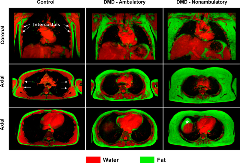
Objective: Duchenne muscular dystrophy (DMD) is characterized by damage to muscles including the muscles involved in respiration. Dystrophic muscles become weak and infiltrated with fatty tissue, resulting in progressive respiratory impairment. The objective of this study was to assess respiratory muscle quality and function in DMD using magnetic resonance imaging and to determine the relationship to clinical respiratory function.

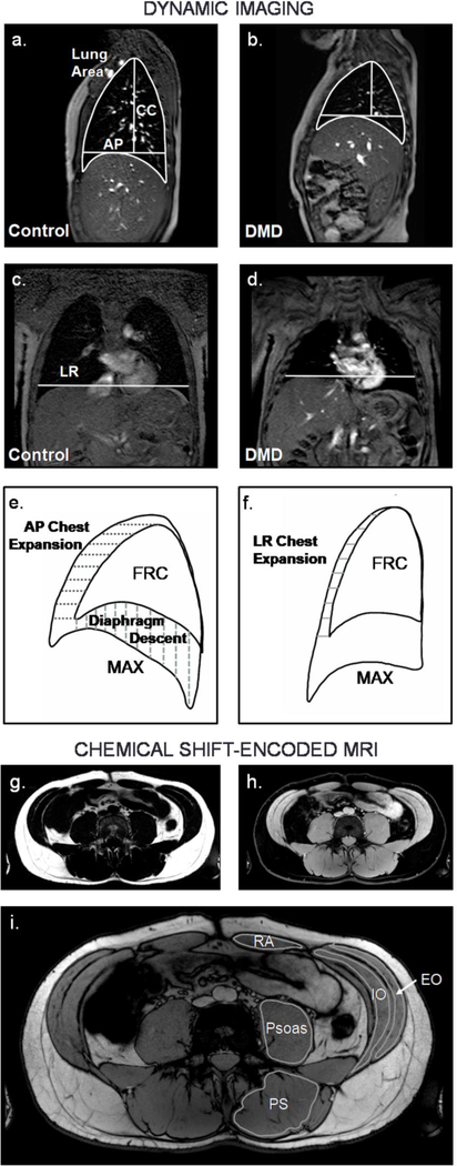
Methods: Individuals with DMD (n = 36) and unaffected controls (n = 12) participated in this cross sectional magnetic resonance imaging study. Participants underwent dynamic imaging of the thorax to assess diaphragm and chest wall mobility and chemical shift-encoded imaging of the chest and abdomen to determine fatty infiltration of the accessory respiratory muscles. Additionally, clinical pulmonary function measures were obtained.

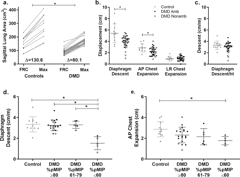
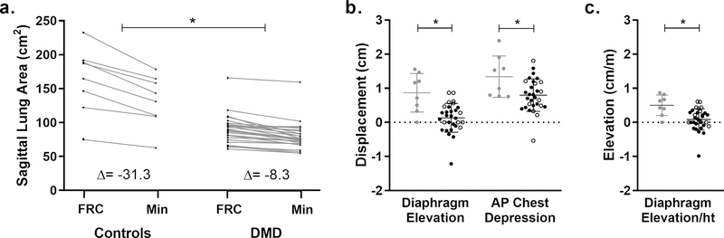
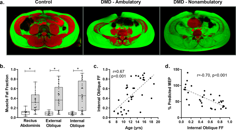
Results: Thoracic cavity area was decreased in individuals with DMD compared to controls during tidal and maximal breathing. Individuals with DMD had reduced chest wall movement in the anterior-posterior direction during maximal inspirations and expirations, but diaphragm descent during maximal inspirations (normalized to height) was only decreased in a subset of individuals with maximal inspiratory pressures less than 60% predicted. Muscle fat fraction was elevated in all three expiratory muscles assessed (p < 0.001), and the degree of fatty infiltration correlated with percent predicted maximal expiratory pressures (r = - 0.70, p < 0.001). The intercostal muscles demonstrated minimal visible fatty infiltration; however, this analysis was qualitative and resolution limited.

Interpretation: This magnetic resonance imaging investigation of diaphragm movement, chest wall movement, and accessory respiratory muscle fatty infiltration provides new insights into the relationship between disease progression and clinical respiratory function.

Keywords: Diaphragm; Dixon imaging; MRI; Neuromuscular disease; Pulmonary.


Conflict of interest statement

Conflicts of Interest: On behalf of all authors, the corresponding author states that there is no conflict of interest

Figures

Fig 1
Fig 1. Representative dynamic MRIs, analysis parameters, and chemical shift-encoded MRIs.
Dynamic imaging was performed in the sagittal plane (a-b) and coronal plane (c-d) (Control = 12.8 yrs old, individual with DMD = 12.3 yrs old). Lung size measurements included sagittal plane lung area, anteriorposterior (AP) chest diameter measured at the level of the top of the diaphragm, craniocaudal (CC) length measured from the apex of the lung to the AP chest diameter line, and left-right (LR) chest width measured at the level of the top of the right diaphragm dome. e) Diaphragm movement after a maximal inspiration or expiration was quantified in the sagittal image, and chest movement was quantified in the sagittal and f) coronal images. CSE MRIs were acquired at the chest and abdomen. They were reconstructed to produce fat g) and water h) images, and FF was quantified for the abdominal muscles indicated in the CSE out-of-phase image in i). FRC = functional residual capacity, MAX = maximal inspiration, RA = rectus abdominis, EO = external oblique, IO = internal oblique, PS = paraspinals, CSE = chemical shift-encoded, FF = fat fraction
Fig 2
Fig 2. Lung area increase, diaphragm descent, and chest expansion during a maximal inspiration.
a) Unaffected controls had larger sagittal plane lung areas at functional residual capacity (FRC) and at maximal inspiration (max) than participants with DMD, and they also had larger increases in lung area during maximal inspirations (p=0.003). b) Additionally, controls (gray circles) had larger diaphragm descent (p=0.015) and anterior-posterior (AP) chest expansion (p=0.015) compared to individuals with DMD (ambulatory = closed black circles, nonambulatory = open black circles), but there was no difference in left-right (LR) chest expansion. c) When diaphragm descent was normalized to height, differences were no longer significant between groups. d) However, the subgroup of participants with DMD with percent predicted MIP (%pMIP) ≤60 had significantly reduced diaphragm descent normalized to height and e) AP chest expansion compared to controls. *Indicates p<0.05
Fig 3
Fig 3. Lung area decrease, diaphragm elevation, and chest depression during a maximal expiration.
a) In addition to having larger sagittal plane lung areas after a maximal expiration, control participants had larger absolute decreases in lung area during maximal expirations (min) compared to participants with DMD (p<0.001). b) Diaphragm elevation and anterior-posterior (AP) chest depression were also significantly reduced in participants with DMD (ambulatory = closed black circles, nonambulatory = open black circles)compared to controls (gray circles). Several participants with DMD had paradoxical diaphragm movement during maximal expirations, and one participant (not the same outlier for diaphragm movement) had paradoxical chest movement. These cases correspond with the negative values in (b). c) Diaphragm elevation was significantly reduced in DMD compared to controls even after normalization to height (ht). *Indicates p<0.05
Fig 4
Fig 4. Expiratory muscle fat fraction.
a) Fat ( green) an d water (red) fusion images of the abdominal and expiratory muscles of a 16 yr old unaffected control, an ambulatory 13 yr old with DMD, and a nonambulatory 16 yr old with DMD. Significant fatty infiltration of the expiratory muscles is visible in the individuals with DMD, and early involvement of the internal oblique was consistently seen even in ambulatory participants. b) Fat fraction (FF) was elevated in DMD compared to controls for each expiratory muscle analyzed. The rectus abdominis was, on average, the least affected expiratory muscle, while the internal oblique was the most affected. c) Internal oblique FF increased with increasing age (r=0.67, p<0.001). d) Internal oblique FF was significantly correlated with percent predicted maximal expiratory pressure (%pMEP) (r=−0.70, p<0.001). *Indicates p<0.05
Fig 5
Fig 5. Fat-water fusion images of the chest.
Fat (green) and water (red) images of the chest were acquired in the coronal and axial planes and overlaid to produce fusion images for a 17 yr old unaffected control, an ambulatory 12 yr old with DMD, and a nonambulatory 12 yr old with DMD. In the individuals with DMD, fatty infiltration is visible in the muscles of the chest. The serratus anterior, which is visible overlying the ribs in the coronal image of the control participant is completely infiltrated with fat in the nonambulatory individual with DMD. However, the intercostal muscles, which are accessory respiratory muscles located between the ribs and beneath the serratus anterior, demonstrate less involvement than the other chest muscles. (Note: In the nonambulatory participant, a pocket of fatty tissue, denoted by an asterisk, is visible anterior to the liver and represents true fat rather than a fat-water switching artifact)
Fig 6
Fig 6. Respiratory MR biomarkers, LE MR biomarkers, and ambulatory status.
a) To assess the relationship between disease progression in the expiratory muscles and other skeletal muscles, expiratory muscle FF, lower trunk muscle (psoas and PS) FF, and lower extremity muscle (VL and SOL) FF were compared. White cells represent missing data. The relationship between FF of the different muscles was generally consistent with lower fatty infiltration of the expiratory muscles being associated with lower fatty infiltration of the trunk and lower extremity muscles. However, some individuals stand out as having one muscle which is more severely affected than the others. For example, participant 11 has a higher paraspinal FF than would be expected given the FF in his other muscles, and participant 16 has a higher IO FF than may be expected. b) During maximal inspirations, nonambulatory individuals had significantly smaller increases in sagittal lung area normalized to height, compared to ambulatory individuals (p=0.046). c) Nonambulatory participants also had significantly higher expiratory muscle FF compared to ambulatory participants (p<0.001 for all comparisons). (EO = external oblique, FF = fat fraction, IO = internal oblique, PS = paraspinals, RA = rectus abdominis, SOL = soleus, VL = vastus lateralis)

References

    1. Mendell JR, Shilling C, Leslie ND, et al. (2012) Evidence-based path to newborn screening for Duchenne muscular dystrophy. Ann Neurol 71:304–313. 10.1002/ana.23528 - DOI - PubMed
    1. Moat SJ, Bradley DM, Salmon R, et al. (2013) Newborn bloodspot screening for Duchenne muscular dystrophy: 21 years experience in Wales (UK). Eur J Hum Genet 21:1049. - PMC - PubMed
    1. Hoffman EP, Brown RH Jr., Kunkel LM (1987) Dystrophin: The protein product of the Duchenne muscular dystrophy locus. Cell 51:919–928. 10.1016/0092-8674(87)90579-4 - DOI - PubMed
    1. Peverelli L, Testolin S, Villa L, et al. (2015) Histologic muscular history in steroid-treated and untreated patients with Duchenne dystrophy. Neurology 85:1886–1893 - PMC - PubMed
    1. Petrof BJ, Shrager JB, Stedman HH, et al. (1993) Dystrophin protects the sarcolemma from stresses developed during muscle contraction. Proc Natl Acad Sci 90:3710–3714 - PMC - PubMed

MeSH terms

LinkOut - more resources