. 2019 Nov 21;101(5):1001-1017.
doi: 10.1093/biolre/ioz139.

Yes-associated protein 1 is required for proliferation and function of bovine granulosa cells in vitro†

Affiliations

Yes-associated protein 1 is required for proliferation and function of bovine granulosa cells in vitro†

Michele R Plewes et al. Biol Reprod. .

Abstract

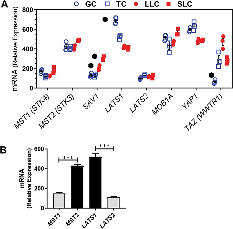
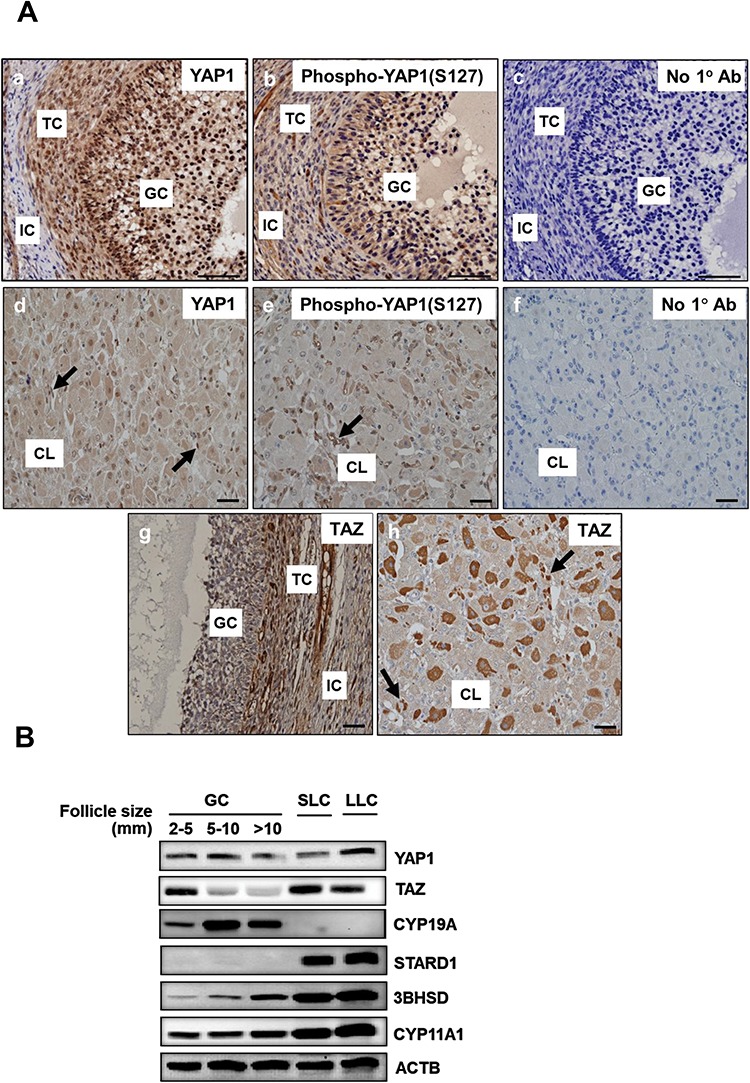
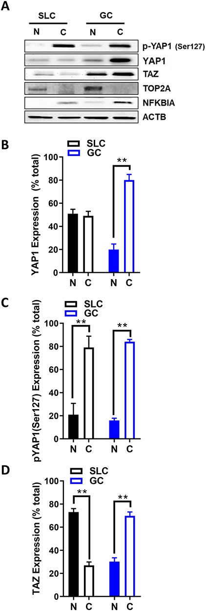
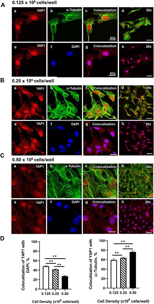
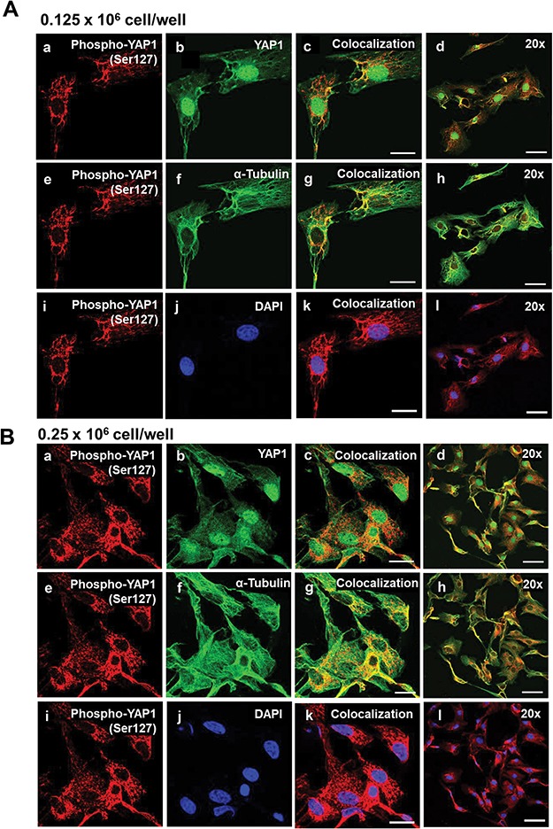
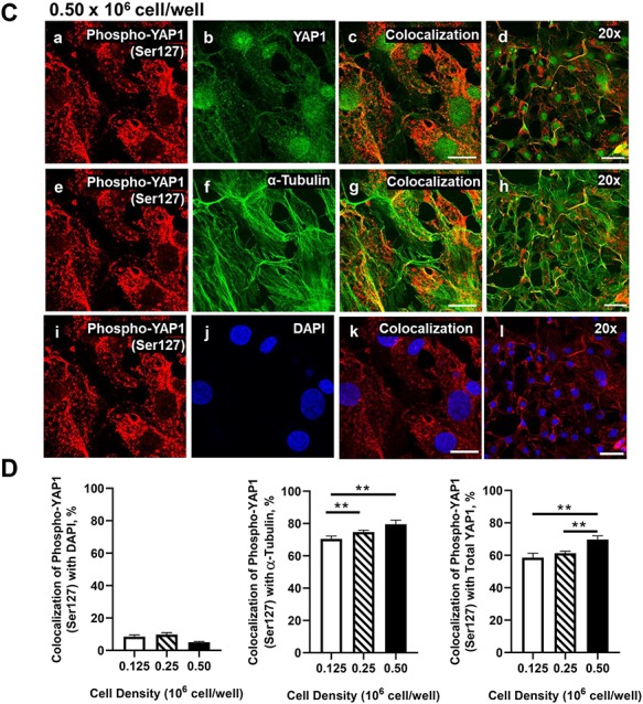
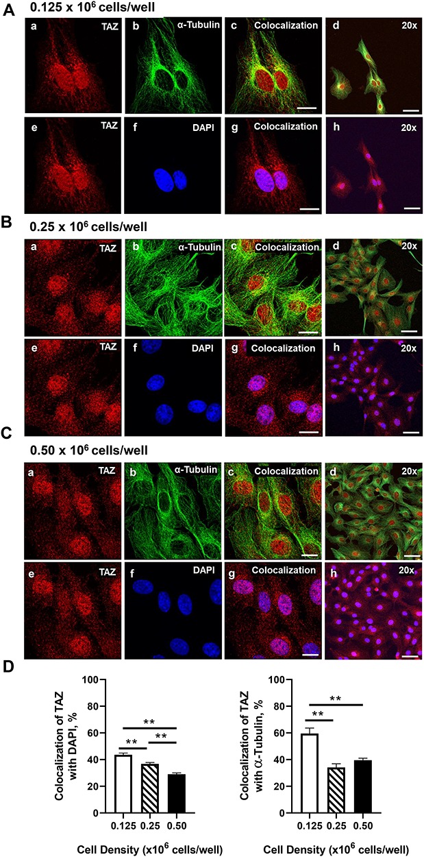
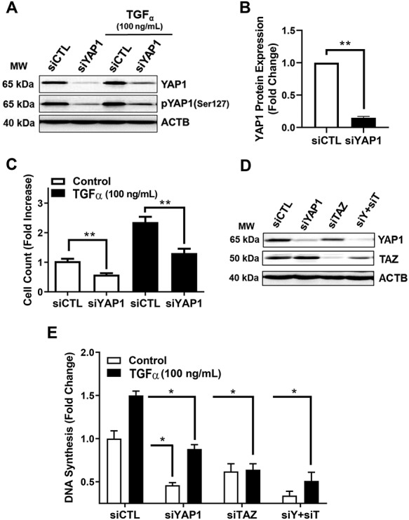
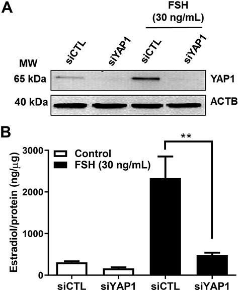
Yes-associated protein 1 (YAP1) is a major component of the Hippo signaling pathway. Although the exact extracellular signals that control the Hippo pathway are currently unknown, increasing evidence supports a critical role for the Hippo pathway in embryonic development, regulation of organ size, and carcinogenesis. Granulosa cells (GCs) within the ovarian follicle proliferate and produce steroids and growth factors, which facilitate the growth of follicle and maturation of the oocyte. We hypothesize that YAP1 plays a role in proliferation and estrogen secretion of GCs. In the current study, we examined the expression of the Hippo signaling pathway in bovine ovaries and determined whether it was important for GC proliferation and estrogen production. Mammalian STE20-like protein kinase 1 (MST1) and large tumor suppressor kinase 2 (LATS2) were identified as prominent upstream components of the Hippo pathway expressed in granulosa and theca cells of the follicle and large and small cells of the corpus luteum. Immunohistochemistry revealed that YAP1 was localized to the nucleus of growing follicles. In vitro, nuclear localization of the downstream Hippo signaling effector proteins YAP1 and transcriptional co-activator with PDZ-binding motif (TAZ) was inversely correlated with GC density, with greater nuclear localization under conditions of low cell density. Treatment with verteporfin and siRNA targeting YAP1 or TAZ revealed a critical role for these transcriptional co-activators in GC proliferation. Furthermore, knockdown of YAP1 in GCs inhibited follicle-stimulating hormone (FSH)-induced estradiol biosynthesis. The data indicate that Hippo pathway transcription co-activators YAP1/TAZ play an important role in GC proliferation and estradiol synthesis, two processes necessary for maintaining normal follicle development.

Keywords: Hippo signaling; Yes-associated protein 1; bovine; follicular development; granulosa cells; proliferation; steroidogenesis.


Figures

Figure 1
Figure 1
Expression of Hippo signaling family members in the bovine ovary. Microarray analysis was used to determine the expression of Hippo signaling family members in bovine follicular (theca and granulosa) and luteal (small and large) cells. (A) Microarray analysis of Hippo signaling family members in bovine GCs (n = 4; open blue circle), TCs (n = 3; open blue box), SLCs (n = 3; closed red box) and LLCs (n = 3; closed red circle). Relative mRNA units are shown as symbols with means ± SEM shown as black lines, in some cases, the black lines are obscured by the symbols. Significant differences were identified as changes greater than 1.5-fold and supported by unpaired t-tests with P < 0.01. *Significant difference between GC and LLC or TC and SLC. Serine/threonine kinase 4 (STK4; MST1); serine/threonine kinase 3 (STK3, MST2); salvador family WW domain containing protein 1 (SAV1); large tumor suppressor kinase 1 (LATS1); large tumor suppressor kinase 2 (LATS2); MOB kinase activator 1A (MOB1A); Yes-associated protein 1 (YAP1); WW domain containing transcription regulator 1 (TAZ; WWTR1); and beta-actin (ACTB). (B) Comparison of the expression of MST1 versus MST2 and LATS1 versus LATS2. Levels of mRNA for each transcript in GC, TC, SLC, and LLC were pooled and analyzed as a single group. Data are means ± SEM. Differences in means in MST1 versus MST2 and LATS1 versus LATS2 were compared by t-test. ***P < 0.001.
Figure 2
Figure 2
Localization of Hippo signaling family members in the bovine ovary. Immunohistochemistry and western blotting were used to determine the expression and localization of YAP1, phospho-YAP1(Ser127), and TAZ in bovine follicles and the corpus luteum. (A) Representative immunohistochemistry micrographs showing expression of YAP1 in GCs and TCs (b) and corpus luteum (d), phospho-YAP1(Ser127) in GC and TC (c) and corpus luteum (e); and TAZ in GC and TC (g) and corpus luteum (h). Micron bar represents 1 mm, negative controls (a and f). Large arrows point to luteal endothelial cells. (B) Western blot analysis of the expression of YAP1 and TAZ in GC from follicles of increasing size (2–5, 5–10, >10 mm) and enriched SLCs and LLCs. Expression of aromatase (CYP19A1), steroidogenic acute regulatory protein (STARD1), 3beta-Hydroxysteroid dehydrogenase (3BHSD), Cholesterol side-chain cleavage enzyme (CYP11A1), and Beta-actin (ACTB; loading control) are shown.
Figure 3
Figure 3
Expression of YAP1 and WW domain-containing transcription regulator 1 (TAZ; WWTR1) in the bovine ovary. Western blotting was used to determine the localization of YAP1, phospho-YAP1(Ser127), and TAZ in bovine follicles and the corpus luteum. (A) Representative western blots of YAP1, phospho-YAP1(Ser127) and TAZ in cytoplasmic and nuclear fractions obtained from cultured GC and enriched SLC. Nuclear protein: DNA topoisomerase II alpha (TOP2A); cytosolic protein: nuclear factor of kappa light polypeptide gene enhancer in B-cells 1 (NFKB1A); beta-actin (ACTB; loading control). (B–D) Densitometry of YAP1, phospho-YAP1(Ser127), and TAZ expression in nuclear and cytoplasmic fractions. Data represent the percentage of each protein within each fraction. Bars are means ± SEM, n = 5 experiments. **P < 0.01.
Figure 4
Figure 4
Effects of cell density on nuclear localization of YAP1 in bovine GCs. Granulosa cells were seeded overnight on coverslips in six-well dishes at increasing cell densities from 0.125–0.50 × 106 cells/well. (A) Representative micrograph of GC plated at 0.125 × 106 cells/well; YAP1 (a), alpha-tubulin (b), colocalization of YAP1 and alpha-tubulin (c), 20× magnification of colocalization of YAP1 and alpha-tubulin (d), YAP1 (e), DAPI (e), colocalization of YAP1 and DAPI (g), 20× magnification of colocalization of YAP1 and alpha-tubulin (h). (B) Representative micrograph of GC plated at 0.25 × 106 cells/well. (C) Representative micrograph of GC plated at 0.50 × 106 cells/well. (D) Quantitative analysis of colocalization of YAP1 with alpha-tubulin and DAPI. Data are represented as means ± SEM, n = 3 experiments. **Significant difference as compared to 0.125 × 106 cells/well, P < 0.05. Micron bar represents 20 μm (63×) and 50 μm (20×).
Figure 5
Figure 5
Effects of cell density on nuclear localization of phosphorylated YAP1 (Ser127) in bovine GCs. Granulosa cells were seeded overnight on coverslips in six-well dishes at increasing cell densities from 0.125 to 0.50 × 106 cells/well. (A) Representative micrograph of GC plated at 0.125 × 106 cells/well; phosphorylated Yes-associated protein (p-YAP1 (Ser127)) (a), YAP1 (b), colocalization of p-YAP1 (Ser127) and YAP1 (c), 20x magnification of colocalization of p-YAP1 (Ser127) and YAP1 (d), p-YAP1 (Ser127) (e), alpha-tubulin (f), colocalization of p-YAP1 (Ser127) and alpha-tubulin (g), 20× magnification of colocalization of p-YAP1 (Ser127) and alpha-tubulin (h), p-YAP1 (Ser127) (i), DAPI (j), colocalization of p-YAP1 (Ser127) and DAPI (k), 20× magnification of colocalization of p-YAP1 (Ser127) and alpha-tubulin (l). (B) Representative micrograph of GC plated at 0.25 × 106 cells/well. (C) Representative micrograph of GC plated at 0.50 × 106 cells/well. (D) Quantitative analysis of colocalization of p-YAP1 with YAP1, alpha-tubulin, and DAPI. Data are represented as means ± SEM, n = 3 experiments. **Significant difference as compared to 0.125 × 106 cells/well, P < 0.05. Micron bar represents 20 μm (63×) and 50 μm (20×).
Figure 5
Figure 5
(Continued).
Figure 6
Figure 6
Effects of cell density on nuclear localization of TAZ in bovine GCs. Granulosa cells were seeded overnight on coverslips in 6-well dishes at increasing cell densities from 0.125–0.50 × 106 cells/well. (A) Representative micrograph of GC plated at 0.125 × 106 cells/well; WW domain-containing transcription regulator 1 (TAZ; WWTR1) (a), alpha-tubulin (b), colocalization of TAZ and alpha-tubulin (c), 20× magnification of colocalization of TAZ and alpha-tubulin (d), TAZ (e), DAPI (e), colocalization of TAZ and DAPI (g), 20× magnification of colocalization of TAZ and alpha-tubulin (h). (B) Representative micrograph of GC plated at 0.25 × 106 cells/well. (C) Representative micrograph of GC plated at 0.50 × 106 cells/well. (D) Quantitative analysis of colocalization of TAZ with alpha-tubulin and DAPI. Data are represented as means ± SEM, n = 3 experiments. **Significant difference as compared to 0.125 × 106 cells/well, P < 0.05. Micron bar represents 20 μm (63×) and 50 μm (20×).
Figure 7
Figure 7
Effects of the YAP1 inhibitor verteporfin on GC proliferation. Bovine GC were plated at a cell density of 80 × 103 cells/well in 12-well dishes and treated with increasing concentrations of verteporfin (0–5 μM). Cell proliferation was determined as described in the Methods. (A) Representative western blot analysis showing Cyclin D1 expression in cells treated with follicle-stimulating hormone (30 ng/ml; FSH) or transforming growth factor-alpha (100 ng/ml; TGFα) in the presence or absence of verteporfin (5 μM). (B) Densitometry of cyclin D1 expression in cells treated with follicle-stimulating hormone (30 ng/mL; FSH) or transforming growth factor-alpha (100 ng/ml; TGFα). (C) DNA synthesis in cells treated with increasing concentrations of verteporfin (0, 1, 2.5. and 5 μM) following treatment of TGFα (100 ng/ml). Data are represented as means ± SEM (n = 4) of the average fold change from control in each experiment. **Significant difference as compared to control, P < 0.05.
Figure 8
Figure 8
Effects of siRNA-mediated knockdown of YAP1 and TAZ on TGFα-induced proliferation in GCs. YAP1 and TAZ mRNA were silenced using siYAP1 and siTAZ in bovine GCs. Following knockdown, GCs were treated with or without transforming growth factor-alpha (50 ng/mL; TGFα) to promote cell proliferation. (A) Representative western blot showing phospho-YAP1(Ser127)  and   YAP1   protein   expression  in  siGlo  (siCTL)  or  siYAP1 knockdown GCs, following treatment with or without TGFα. (B) Densitometric analyses of YAP1 protein expression obtained from siCTL (open bars) and siYAP1 (closed bars). Bars represent means ± SEM, n = 5. (C) Cell counts following knockdown of YAP1 cells and treatment with TGFα (closed bars) or without TGFα (open bars). Bars are means ± SEM, n = 3. (D) Representative western blot showing YAP1 and TAZ protein expression in GCs following 48 h treatment with siCTL, siYAP1, siTAZ, or a combination of siYAP1 and siTAZ (siY + siT). Beta-actin (ACTB; loading control). (E) Quantitative analysis showing DNA synthesis for siCTL, siYAP1, siTAZ, or siY + siT knockdown cells treated with control (open bars) or TGFα (black bars). Data are represented as means ± SEM (n = 3) of the average fold change from control in each experiment. **Significant differences between treatment groups, as compared to siCTL, P < 0.05.
Figure 9
Figure 9
Yes-associated protein 1 is required for FSH-induced estradiol production in bovine GCs. Granulosa cells were treated with siGlo (siCTL) or siYAP1 and then treated without (control) or FSH (30 ng/ml) for 48 h prior to analysis of estradiol production. (A) Representative western blot showing YAP1 protein expression. Beta-actin (ACTB; loading control). (B) Quantitative ELISA analysis of estradiol in the culture medium. Data are represented as means ± SEM, n = 3. **Significant differences between treatment groups, as compared to siCTL, P < 0.05.

References

    1. Williams CJ. Erickson GF. Morphology and Physiology of the Ovary. [Updated 2012. Jan 30]. In: Feingold KR, Anawalt B, Boyce A, et al., editors. Endotext [Internet]. South Dartmouth (MA): MDText.com, Inc.; 2000-. Available from: https://www.ncbi.nlm.nih.gov/books/NBK278951/ - PubMed
    1. Kranc W, Budna J, Kahan R, Chachuła A, Bryja A, Ciesiółka S, Borys S, Antosik M, Bukowska D, Brussow K. Molecular basis of growth, proliferation, and differentiation of mammalian follicular granulosa cells. J Biol Regul Homeost Agents 2017; 31:1–8. - PubMed
    1. Fitzpatrick SL, Richards JS. Regulation of the rat aromatase gene in ovarian granulosa cells and R2C Leydig cells. J Steroid Biochem Mol Biol 1993; 44:429–433. - PubMed
    1. Robker RL, Richards JS. Hormone-induced proliferation and differentiation of granulosa cells: a coordinated balance of the cell cycle regulators cyclin D2 and p27Kip1. Mol Endocrinol 1998; 12:924–940v. - PubMed
    1. Regan SL, Knight PG, Yovich JL, Leung Y, Arfuso F, Dharmarajan A. Granulosa cell apoptosis in the ovarian follicle—a changing view. Front Endocrinol 2018; 9:61. - PMC - PubMed

Publication types

MeSH terms