Case Reports
. 2019 Sep;7(9):e818.
doi: 10.1002/mgg3.818. Epub 2019 Jul 27.

TASP1 mutation in a female with craniofacial anomalies, anterior segment dysgenesis, congenital immunodeficiency and macrocytic anemia

Affiliations
Case Reports

TASP1 mutation in a female with craniofacial anomalies, anterior segment dysgenesis, congenital immunodeficiency and macrocytic anemia

Daniel M Balkin et al. Mol Genet Genomic Med. 2019 Sep.

Abstract

Background: Threonine Aspartase 1 (Taspase 1) is a highly conserved site-specific protease whose substrates are broad-acting nuclear transcription factors that govern diverse biological programs, such as organogenesis, oncogenesis, and tumor progression. To date, no single base pair mutations in Taspase 1 have been implicated in human disease.

Methods: A female infant with a new pattern of diagnostic abnormalities was identified, including severe craniofacial anomalies, anterior and posterior segment dysgenesis, immunodeficiency, and macrocytic anemia. Trio-based whole exome sequencing was performed to identify disease-causing variants.

Results: Whole exome sequencing revealed a normal female karyotype (46,XX) without increased regions of homozygosity. The proband was heterozygous for a de novo missense variant, c.1027G>A predicting p.(Val343Met), in the TASP1 gene (NM_017714.2). This variant has not been observed in population databases and is predicted to be deleterious.

Conclusion: One human patient has been reported previously with a large TASP1 deletion and substantial evidence exists regarding the role of several known Taspase 1 substrates in human craniofacial and hematopoietic disorders. Moreover, Taspase 1 deficiency in mice results in craniofacial, ophthalmological and structural brain defects. Taken together, there exists substantial evidence to conclude that the TASP1 variant, p.(Val343Met), is pathogenic in this patient.

Keywords: Taspase 1; anterior segment dysgenesis; congenital immunodeficiency; craniofacial anomaly; macrocytic anemia.


Conflict of interest statement

None declared.

Figures
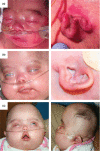
Figure 1
Figure 1
Clinical photographs of a female patient with a de novo missense variant in the TASP1 gene. Frontal (left) and lateral (right) views at 2 days (a), 2 months, note involution of the congenital hemangioma (b) and 17 months of age (c)
Figure 2
Figure 2
Pre‐ and postoperative cross‐sectional imaging. Magnetic resonance imaging at 3 days of age (a–d). Axial diffusion‐weighted image revealing multifocal ischemia involving the left temporal lobe (star) with additional areas of microhemorrhage (triangle) (a). Axial multi‐planar reconstruction illustrating left temporal lobe extra‐axial hemorrhagic collection (star) and anomalous course of the bilateral optic nerves (triangle) (b). Axial multiplanar reconstruction (b) and sagittal T2‐weighted (c) images with notable prominent and oblong optic globes, as well as delayed parenchymal sulcation (c, star). Sagittal T1‐weighted image demonstrating thinning of the corpus callosum  (star) and absence of a Chiari malformation (triangle) (d). Pre‐operative computed tomography at 14‐months of age (e–g). Axial image revealing complete failure of ossification of the greater wing of the sphenoid bones (triangle) and bilateral temporal squamosa (e). Axial image demonstrating hypo‐ossification of the skull base (f). Three dimensional reconstruction with large anterior fontanelle (frontal, left) and absence of temporal bone squamosa (lateral, right). Postoperative computed tomography three dimensional reconstruction at 17‐months of age demonstrating sliding interdigitating parietal bone grafts (lateral, right) and a bandeau orbital advancement with partial reduction of the anterior fontanelle with frontal and temporal bone grafts (frontal, left) (h)
Figure 3
Figure 3
TASP1, downsteam targets and human conditions. The 50 kD Taspase 1 alpha‐beta proenzyme undergoes intramolecular auto proteolysis generating the active alpha‐beta heterodimer (a). MLL/KMT2A is cleaved by TASP1, resulting in active histone H3 methylation and downstream transcriptional changes (b, left). TASP1 also uses MLL2/KMT2D as substrate rending active histone modification (b, right). The transcription factor TFIIA (GTF2A1), an additional TASP1 substrate, is one of several basal transcription factors required for all transcriptional events that use RNA polymerase II. Following Taspase 1‐mediated cleavage, TFIIA is susceptible to proteosome‐mediated degradation (c, left). However, when TASP1 is absent, there is an upregulated expression of TFIIA target genes (CDKN1A/P21CIP1 and CDKN2A/P16INK4A/P14ARF), which impacts craniofacial development (c, right). Of note, red text indicates human pathologies resulting from mutations in the indicated genes (TASP1, MLL/KMT2A, and MLL2/KMT2D)
Figure 4
Figure 4
Analysis of p.Val343Met Mutation. Amino acid sequence alignment across various species with colors denoting conservation scores (dark pink, highly conserved; dark blue, highly variable) (a, arrow indicates valine 343). Crystal structure of the TASP1 heterotetramer (b, left, ball and stick representation; right, space filling model color coded to conservation scores, box containing valine 343). Close up view of valine 343 with conservation color analysis (c, star indicates pink valine 343)

References

    1. Adzhubei, I. A. , Schmidt, S. , Peshkin, L. , Ramensky, V. E. , Gerasimova, A. , Bork, P. , … Sunyaev, S. R. (2010). A method and server for predicting damaging missense mutations. Nature Methods, 7(4), 248–249. 10.1038/nmeth0410-248 - DOI - PMC - PubMed
    1. Armstrong, S. A. , Staunton, J. E. , Silverman, L. B. , Pieters, R. , den Boer, M. L. , Minden, M. D. , … Korsmeyer, S. J. (2002). MLL translocations specify a distinct gene expression profile that distinguishes a unique leukemia. Nature Genetics, 30(1), 41–47. 10.1038/ng765 - DOI - PubMed
    1. Ayton, P. M. , & Cleary, M. L. (2001). Molecular mechanisms of leukemogenesis mediated by MLL fusion proteins. Oncogene, 20(40), 5695–5707. 10.1038/sj.onc.1204639 - DOI - PubMed
    1. Capotosti, F. , Hsieh, J. J. , & Herr, W. (2007). Species selectivity of mixed‐lineage leukemia/trithorax and HCF proteolytic maturation pathways. Molecular and Cellular Biology, 27(20), 7063–7072. 10.1128/MCB.00769-07 - DOI - PMC - PubMed
    1. Chen, C. , Liu, Y. U. , Rappaport, A. R. , Kitzing, T. , Schultz, N. , Zhao, Z. , … Lowe, S. W. (2014). MLL3 is a haploinsufficient 7q tumor suppressor in acute myeloid leukemia. Cancer Cell, 25(5), 652–665. 10.1016/j.ccr.2014.03.016 - DOI - PMC - PubMed

Publication types

MeSH terms

Supplementary concepts