Review
. 2020 Feb 20;48(3):1013-1028.
doi: 10.1093/nar/gkz637.

Hallmarks of ribosomopathies

Affiliations
Review

Hallmarks of ribosomopathies

Kim R Kampen et al. Nucleic Acids Res. .

Abstract

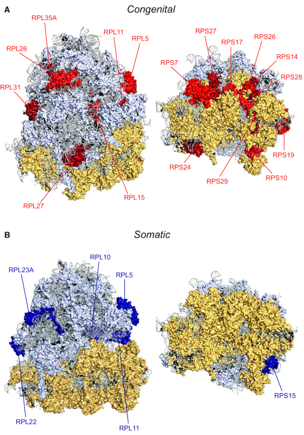
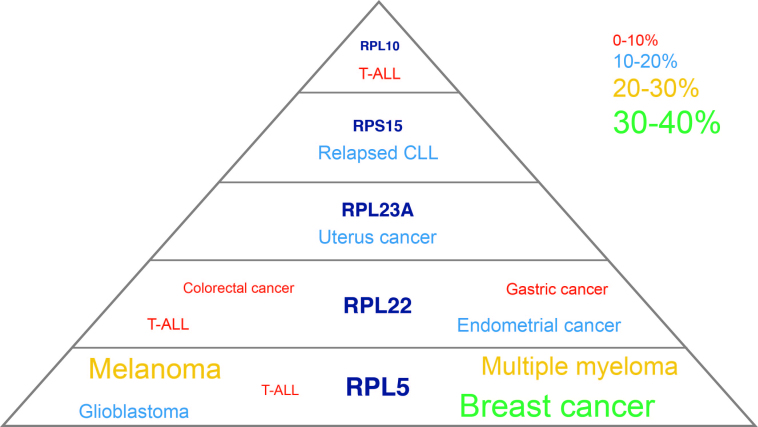
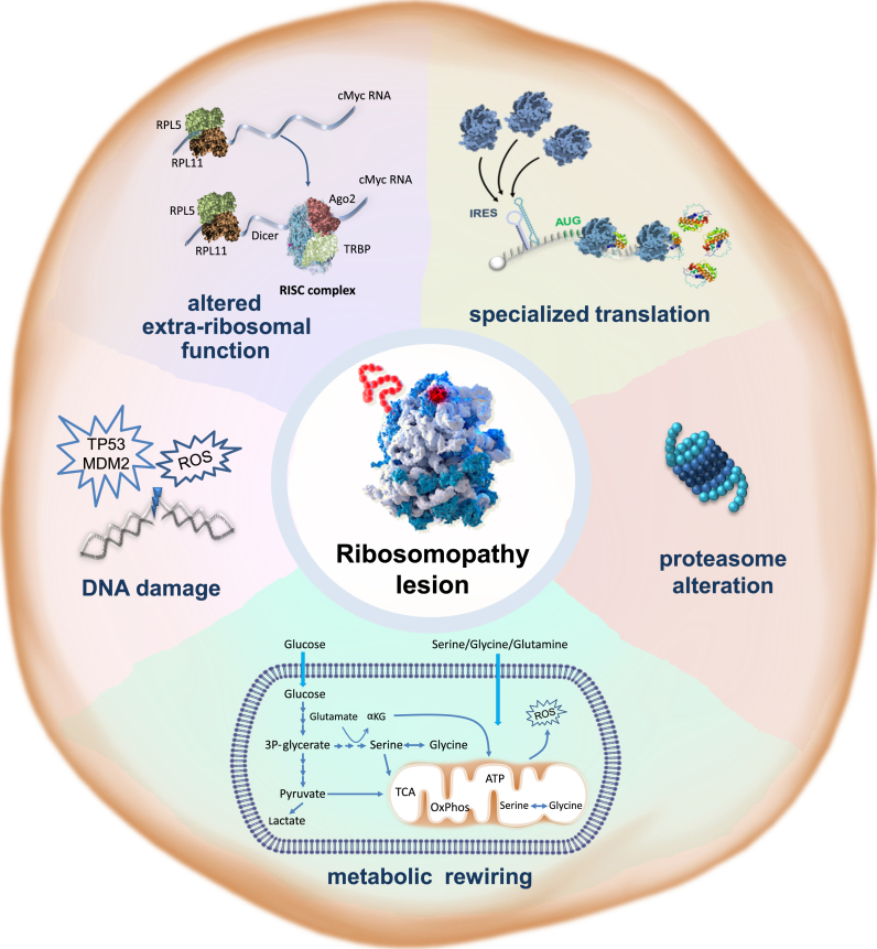
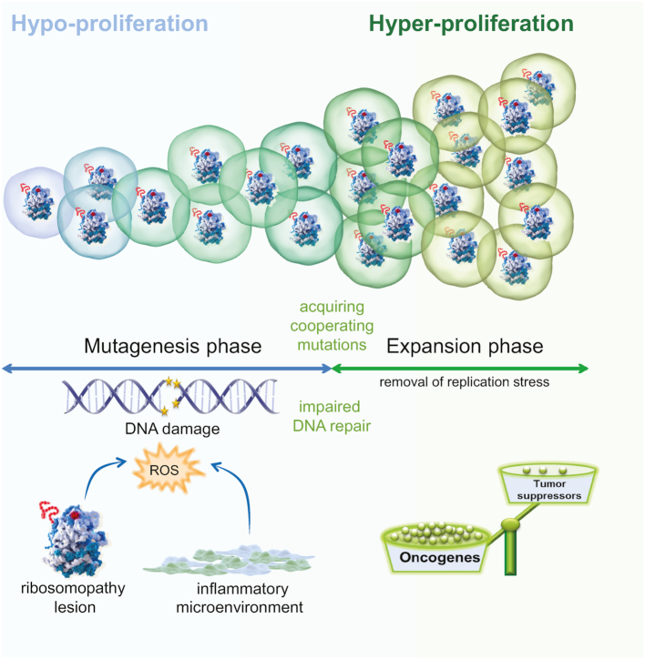
Ribosomopathies are diseases caused by defects in ribosomal constituents or in factors with a role in ribosome assembly. Intriguingly, congenital ribosomopathies display a paradoxical transition from early symptoms due to cellular hypo-proliferation to an elevated cancer risk later in life. Another association between ribosome defects and cancer came into view after the recent discovery of somatic mutations in ribosomal proteins and rDNA copy number changes in a variety of tumor types, giving rise to somatic ribosomopathies. Despite these clear connections between ribosome defects and cancer, the molecular mechanisms by which defects in this essential cellular machinery are oncogenic only start to emerge. In this review, the impact of ribosomal defects on the cellular function and their mechanisms of promoting oncogenesis are described. In particular, we discuss the emerging hallmarks of ribosomopathies such as the appearance of 'onco-ribosomes' that are specialized in translating oncoproteins, dysregulation of translation-independent extra-ribosomal functions of ribosomal proteins, rewired cellular protein and energy metabolism, and extensive oxidative stress leading to DNA damage. We end by integrating these findings in a model that can provide an explanation how ribosomopathies could lead to the transition from hypo- to hyper-proliferation in bone marrow failure syndromes with elevated cancer risk.


Figures

Figure 1.
Figure 1.
Structural model of the ribosome with indication of RPs affected in ribosomopathies. Structural model of the human 80S ribosome with indication of RPs with recurrent mutations and/or deletions in ribosomopathies. The 60S large subunit, 40S small subunit and ribosomal RNA are indicated in yellow, light blue and gray, respectively. In panel (A), RPs with congenital defects are indicated in red. RPs with somatic mutations and deletions are marked in blue in panel (B). Each panel shows two different viewpoints of the solvent side of the ribosome. This figure was generated in PyMOL and is based on the human cryo-EM structure with a resolution of 3.9Å (PDM entry: 6IP5) (140). New RP nomenclature: RPL5 (uL18), RPL10 (uL16), RPL11 (uL5), RPL15 (eL15), RPL22 (eL22), RPL23A (uL23), RPL26 (uL24), RPL27 (eL27), RPL31 (eL31), RPL35A (eL33), RPS7 (eS7), RPS10 (eS10), RPS14 (uS11), RPS15 (uS19), RPS17 (eS17), RPS19 (eS19), RPS24 (eS24), RPS26 (eS26), RPS27 (eS27), RPS28 (eS28), RPS29 (uS14).
Figure 2.
Figure 2.
Scheme illustrating the RP genes with recurrent somatic mutations and/or deletions in cancer. Font sizes in this figure are proportional to incidence of the RP mutations and deletions in the cancer types where they have been described, and incidence percentages of the RP defects are indicated by the color legend; CLL, chronic lymphocytic leukemia; T-ALL, T-cell acute lymphoblastic leukemia.
Figure 3.
Figure 3.
Hallmarks of ribosomopathies. Ribosomopathies are characterized by a collection of cellular phenotypes that promote cancer as summarized in this figure that is inspired by the figure from the landmark ‘Hallmarks of Cancer’ review by Hanahan and Weinberg (11). (i) Ribosomopathy lesions reprogram translation, affecting cellular translation of a subset of hematopoietic and cancer-promoting mRNAs. (ii) Many ribosomopathies display an altered proteasome function, which can lead to stabilization or increased degradation of subsets of proteins, including oncogenes and tumor suppressors. (iii) Ribosomopathies display metabolic rewiring. The implications for translational reprogramming of specific subsets of proteins or supporting an alternative metabolic requirement of RP-defective cells is currently unclear. (iv) Ribosomapthy cells display elevated levels of oxidative DNA damage that can promote acquisition of secondary mutations with a key role in cancer transformation. (v) Lesions can influence the extra-ribosomal function of the mutated RPs.
Figure 4.
Figure 4.
Cellular hypo- to hyper-proliferation transition model. Intracellular oxidative stress caused by alterations in the assembly and/or function of ribosomes or by micro-environmental factors such as inflammation, impairs cell proliferation and promotes DNA damage. Defective DNA repair mechanisms can also induce or further aggravate this DNA damage. After a certain latency, this elevated DNA damage results in the acquisition of rescuing mutations that inhibit ROS production, thereby removing the block on cellular proliferation. At this stage, the expansion phase starts. This phase is then further accelerated by overexpression of oncogenes and/or downregulation of tumor suppressor genes caused by the excessive DNA damage that occurred in the hypoproliferative phase, as well as by reprogramming of the cellular protein translation landscape in the case of RP defects.

References

    1. Warner J.R., Vilardell J., Sohn J.H.. Economics of ribosome biosynthesis. Cold Spring Harb. Symp. Quant. Biol. 2001; 66:567–574. - PubMed
    1. Narla A., Ebert B.L.. Ribosomopathies: human disorders of ribosome dysfunction. Blood. 2010; 115:3196–3205. - PMC - PubMed
    1. Mills E.W., Green R.. Ribosomopathies: there’s strength in numbers. Science. 2017; 358:eaan2755. - PubMed
    1. Sulima S.O., De Keersmaecker K.. Bloody mysteries of ribosomes. Hemasphere. 2018; 2:e95. - PMC - PubMed
    1. Gerst J.E. Pimp my Ribosome: Ribosomal protein paralogs specify translational control. Trends Genet. 2018; 34:832–845. - PubMed

Publication types