. 2019 Dec 1;36(12):2714-2736.
doi: 10.1093/molbev/msz175.

From Root to Tips: Sporulation Evolution and Specialization in Bacillus subtilis and the Intestinal Pathogen Clostridioides difficile

Affiliations

From Root to Tips: Sporulation Evolution and Specialization in Bacillus subtilis and the Intestinal Pathogen Clostridioides difficile

Paula Ramos-Silva et al. Mol Biol Evol. .

Abstract

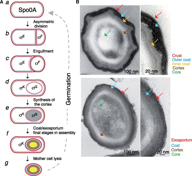
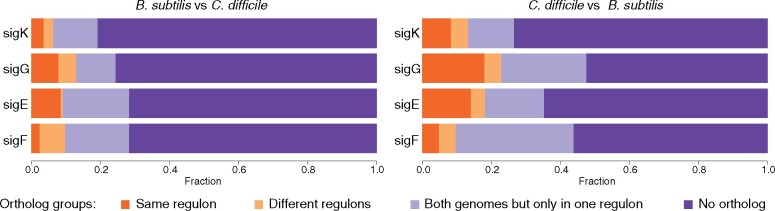
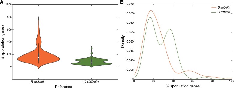
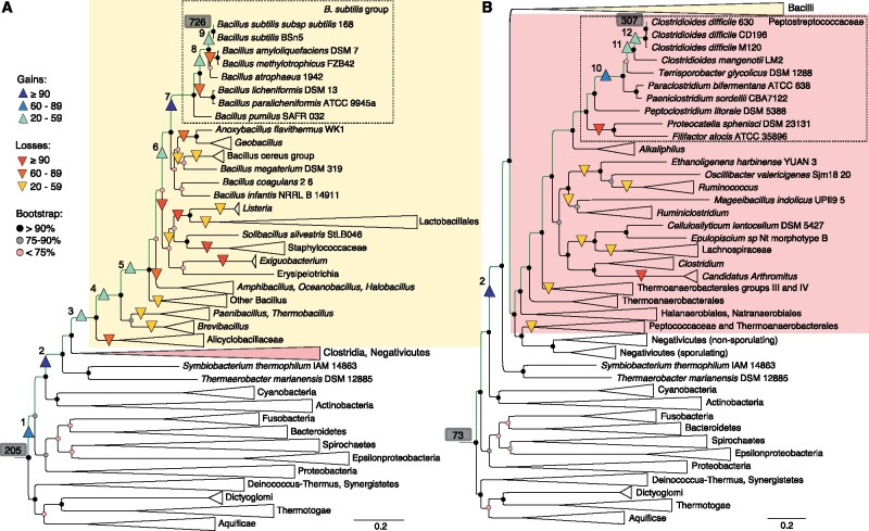
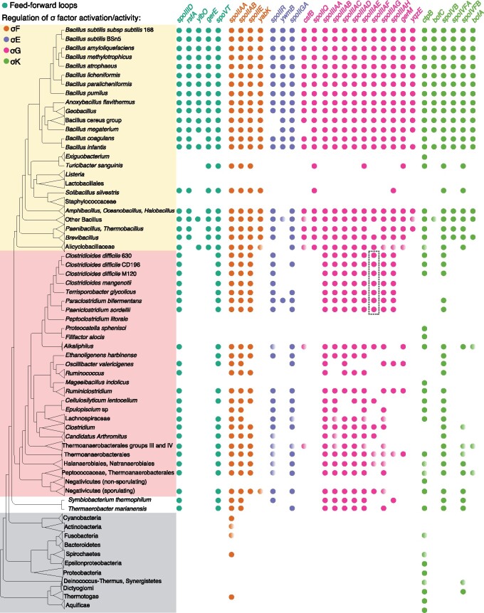
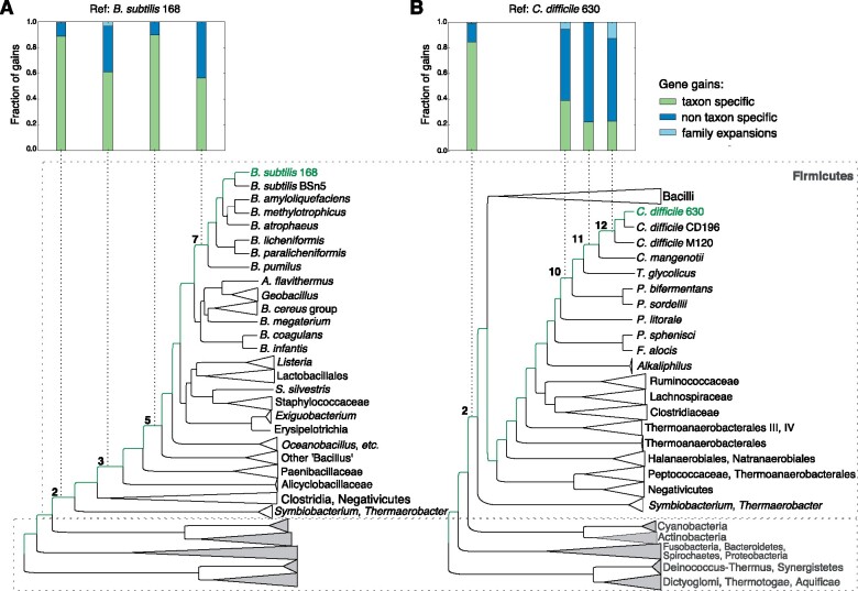
Bacteria of the Firmicutes phylum are able to enter a developmental pathway that culminates with the formation of highly resistant, dormant endospores. Endospores allow environmental persistence, dissemination and for pathogens, are also infection vehicles. In both the model Bacillus subtilis, an aerobic organism, and in the intestinal pathogen Clostridioides difficile, an obligate anaerobe, sporulation mobilizes hundreds of genes. Their expression is coordinated between the forespore and the mother cell, the two cells that participate in the process, and is kept in close register with the course of morphogenesis. The evolutionary mechanisms by which sporulation emerged and evolved in these two species, and more broadly across Firmicutes, remain largely unknown. Here, we trace the origin and evolution of sporulation using the genes known to be involved in the process in B. subtilis and C. difficile, and estimating their gain-loss dynamics in a comprehensive bacterial macroevolutionary framework. We show that sporulation evolution was driven by two major gene gain events, the first at the base of the Firmicutes and the second at the base of the B. subtilis group and within the Peptostreptococcaceae family, which includes C. difficile. We also show that early and late sporulation regulons have been coevolving and that sporulation genes entail greater innovation in B. subtilis with many Bacilli lineage-restricted genes. In contrast, C. difficile more often recruits new sporulation genes by horizontal gene transfer, which reflects both its highly mobile genome, the complexity of the gut microbiota, and an adjustment of sporulation to the gut ecosystem.

Keywords: bacterial genome evolution; horizontal gene transfer; sporulation; taxon-specific genes, Bacillus subtilis, Clostridioides difficile.


Figures

<sc>Fig</sc>. 1.
Fig. 1.
Sporulation stages and spore ultrastructure. (A) Depicts the main stages of sporulation as described for the model organisms B. subtilis. At the onset of the process, the rod-shaped cells (a) divide asymmetrically to produce a larger mother cell and a smaller forespore (the future spore) (b). Asymmetric division involves PG synthesis within the septum. The mother cell then starts to engulf the forespore (c), eventually releasing it as a free protoplast inside its cytoplasm (d). Following engulfment completion, the forespore is no longer in contact with the external medium and is separated from the mother cell by a system of two membranes that derive from the asymmetric division septum, the inner and outer forespore membranes. Following engulfment completion, the forespore becomes visible as a phase dark body inside the mother cell (e). Synthesis of the primordial germ cell wall takes place from the forespore, whereas synthesis of the spore cortex PG layer is a function of the mother cell. Development of full spore refractility coincides with the formation of the cortex. The final stages in the assembly of the spore coat and crust are also represented (f). Finally, the spore is released into the environment through autolysis of the mother cell (g). Spores will resume vegetative growth through the process of germination. Spo0A is represented in predivisional cells; the cell in which the cell type-specific sigma factors are active in relation to the stages of sporulation is also represented. (B) Transmission electron microscopy (TEM) image of a thin cross section of a B. subtilis (top) and C. difficile (bottom) spore. The main spore structures are labeled in the diagram. Note that the crust layer of B. subtilis spores is not visible in the microscopy image, but its position, at the edge of the outer coat, is indicated in the diagram. The panels on the right show a magnification of the spore surface layers. The diagram identifies the main structures or compartments normally seen by TEM.
<sc>Fig</sc>. 2.
Fig. 2.
Clustering of candidate orthologs from a pairwise genome comparison between B. subtilis 168 and C. difficile 630. Only clusters including genes controlled by sporulation-specific sigma factors were selected for further analyses. The fraction of orthologous genes sharing the same sporulation regulon is marked in orange. The fraction of orthologs in different regulons is in yellow. The fraction of genes without orthologs in the regulons but with orthologs in the genome is in light purple. The fraction of genes without orthologs is in dark purple.
<sc>Fig</sc>. 3.
Fig. 3.
Distribution of sporulation genes from B. subtilis and C. difficile across the 258 bacterial genomes sampled from NCBI. Distributions are represented in (A) absolute number of genes and (B) relative percentages. Distributions were tested for unimodality using the Hartigan’s Dip test in R (D = 0.024757, P value = 0.3179 for B. subtilis; D = 0.05604, P value = 7.262e-06 for C. difficile).
<sc>Fig</sc>. 4.
Fig. 4.
Sporulation gene gain and loss events across the bacterial phylogeny for (A) Bacilli and (B) Clostridia. The total number of sporulation genes present at the root and at the referential tips is highlighted in gray. Gain events are numbered from 1 to 12. Multigene maximum likelihood (RAxML) tree inferred from an alignment of 70 orthologs and corresponding bootstraps measures in %. The PROTGAMMALG evolutionary model was used to infer the tree with branch support estimated with 100 bootstrap replicates.
<sc>Fig</sc>. 5.
Fig. 5.
Phylogenetic profile showing the conservation of gene presence in the following functional categories: FFLs (dark green) and regulation of activation and activity of σF (orange), σE (purple), σG (pink), and σK (light green). Conservation of gene presence is based on the homology mapping approach described in supplementary figure S2, Supplementary Material online, averaged for collapsed tips that included more than 1 species (e.g., Ruminiclostridium, Actinobacteria) and visualized with EvolView (Zhang et al. 2012). Full circles indicate gene presence above 50%. Half circles indicate gene presence at 50%. No circle indicates gene presence lower than 50% or total absence. On the top, gene reference sequences, names and functional categories were selected based on the B. subtilis sporulation network. Members of the ortholog cluster spoiIIIAF are inside the rectangle.
<sc>Fig</sc>. 6.
Fig. 6.
Increase in size of sporulation regulons represented from root to tips for (A) B. subtilis and (B) C. difficile. Sporulation regulons have diverse sizes being σE the largest regulon and also the one acquiring the largest number of genes in every evolutionary step. To suppress this effect, the gains per regulon were normalized by size.
<sc>Fig</sc>. 7.
Fig. 7.
Predicted categories of sporulation gene gains in four branches for (A) B. subtilis and (B) C. difficile. Gene gains were considered to be taxon specific (green) when gained at the branch, present in its descendants but not present or gained in other clades. Alternatively, gene gains were considered non taxon specific (dark blue) when gained along a certain branch but also present in other nondescendent clades. The later included genes gained by HGT in both directions (from and to the branch/clade). Family expansions (light blue) were originated by duplication events.
<sc>Fig</sc>. 8.
Fig. 8.
Phylogenetic tree and conserved blocks of CdeA proteins. Orthologous sequences were selected based on the results of the orthology mapping approach and supplemented with orthologs from the cluster ENOG410XVF3 from EggNOG 4.5.1 (Huerta-Cepas, Szklarczyk, et al. 2016). Species are colored by environmental origin/niche. (A) Phylogeny with midpoint rooting and bootstrap probabilities were estimated with RAxML (Stamatakis 2014) using the best protein evolution model (LG + I) selected with ProtTest version 3 (Darriba et al. 2011), sequences were aligned using MAFFT (Katoh and Standley 2013). Protein blocks were drawn with ETE3 (Huerta-Cepas, Serra, et al. 2016). (B) Protein logos showing the two blocks of the conserved domain with four cysteines were estimated with WEBLOGO (Crooks et al. 2004).
<sc>Fig</sc>. 9.
Fig. 9.
Comparative analysis between sporulation proteins SpoIIIAF from Bacillus subtilis and SpoiIIIAF from Clostridioides difficile. (A) Ortholog clusters of SpoIIIAF and SpoiIIIAF (n = 64) were merged and aligned using MAFFT (Katoh and Standley 2013) followed by a principal component analysis as implemented in Jalview (Waterhouse et al. 2009) to estimate sequence divergence between clusters. (B) Selected sequences clustering with SpoIIIAF (black square) and SpoiIIIAF (purple square) were aligned with T-coffee (Notredame et al. 2000). (C) Aligned structures of the monomeric forms of SpoIIIAF85-206 (gray) and SpoiIIIAF84-205 (orange) were determined using Modeler homology-based modeling (Eswar et al. 2006) using the structure of B. subtilis SpoIIIAF (PDB code: 6dcs) as the template and MUSCLE (Edgar 2004) as sequence aligner. Structures and electrostatic charge distributions were visualized with Chimera (Pettersen et al. 2004).

Similar articles

Cited by

References

    1. Abanes-De Mello A, Sun YL, Aung S, Pogliano K. 2002. A cytoskeleton-like role for the bacterial cell wall during engulfment of the Bacillus subtilis forespore. Genes Dev. 1624:3253–3264. - PMC - PubMed
    1. Abecasis AB, Serrano M, Alves R, Quintais L, Pereira-Leal JB, Henriques AO. 2013. A genomic signature and the identification of new sporulation genes. J Bacteriol. 1959:2101–2115. - PMC - PubMed
    1. Abt MC, McKenney PT, Pamer EG. 2016. Clostridium difficile colitis: pathogenesis and host defence. Nat Rev Microbiol. 1410:609–620. - PMC - PubMed
    1. Adams CM, Eckenroth BE, Putnam EE, Doublié S, Shen A. 2013. Structural and functional analysis of the CspB protease required for Clostridium spore germination. PLoS Pathog. 92:e1003165.. - PMC - PubMed
    1. Al-Hinai MA, Jones SW, Papoutsakis ET. 2015. The Clostridium sporulation programs: diversity and preservation of endospore differentiation. Microbiol Mol Biol Rev. 791:19–37. - PMC - PubMed

Publication types

LinkOut - more resources