Observational Study
. 2019 Nov;39(12):1054-1063.
doi: 10.1002/pd.5539. Epub 2019 Aug 7.

Transposition of the great arteries: Fetal pulmonary valve growth and postoperative neo-aortic root dilatation

Affiliations
Observational Study

Transposition of the great arteries: Fetal pulmonary valve growth and postoperative neo-aortic root dilatation

Roel L F van der Palen et al. Prenat Diagn. 2019 Nov.

Abstract

Objectives: Documentation of semilunar valve growth in fetal transposition of the great arteries (TGA) and the relationship between neo-aortic root (NAoR) dilatation, a cause for postoperative reinterventions after the arterial switch operation (ASO), and pulmonary valve (PV) annulus dimensions prenatally.

Methods: This retrospective multicenter observational study included TGA fetuses suitable for ASO. Semilunar valve annuli pre-ASO and NAoR diameters (post-ASO) were measured. Trends in annulus diameters were analyzed using a linear mixed-effects model and compared with normal values. Prenatal semilunar valve Z-scores were correlated with NAoR diameters post-ASO.

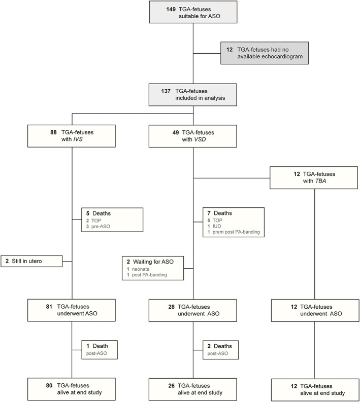
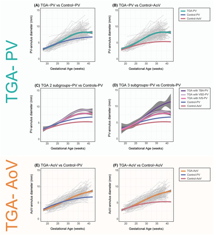
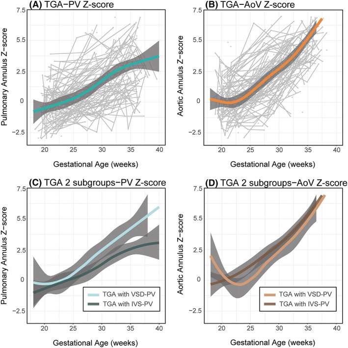
Results: We included 137 TGA fetuses (35.8% with significant ventricular septal defects [VSDs]). One hundred twenty-one underwent ASO. Fetal TGA-PV diameters were significantly larger than control aortic valve (AoV) and PV annuli from 23 and 27 weeks, respectively, especially when a VSD was present. Fetal TGA-AoV annuli were significantly larger than control AoV and PV annuli from 26 and 30 weeks, respectively. Z-scores of fetal TGA-PV and NAoR diameter at last follow-up correlated significantly (P < .001 at 26-30 wk).

Conclusion: Fetal TGA semilunar valve annuli are larger than control annuli, especially when there is a significant VSD. Factors besides postoperative hemodynamics, including fetal anatomy, PV Z-score, prenatal flow, connective tissue properties, and genetics, may influence the risk for late reintervention in these fetuses.


Conflict of interest statement

None declared.

Figures

Figure 1
Figure 1
A, Measurements were made prenatally at the level of the semilunar valve annulus with the valve open. B, Post arterial switch measurements of the aortic root were made in the long axis view with the neo‐aortic valve open; 1 = annulus, 2 = neo‐aortic root, and 3 = sino‐tubular junction. Ao, aorta; LV, left ventricle; PA, pulmonary artery; RV, right ventricle [Colour figure can be viewed at http://wileyonlinelibrary.com]
Figure 2
Figure 2
Flow diagram of included patients. ASO, arterial switch operation; IUD, intra‐uterine death; IVS, intact inter‐ventricular septum; TBA, Taussig‐Bing anatomy; TGA, transposition of the great arteries; TOP, termination of pregnancy; VSD, ventricular septal defect [Colour figure can be viewed at http://wileyonlinelibrary.com]
Figure 3
Figure 3
Average trends in PV annular diameters in TGA fetuses (TGA‐PV): (A) versus trends in controls PV annular diameter (Control‐PV); (B) versus AoV annular diameter (Control‐AoV); (C) TGA with an IVS (TGA with IVS‐PV) and a VSD (TGA with VSD‐PV); and (D) with the TGA‐VSD fetuses further stratified on the basis of a TBA or non‐TBA anatomy (TGA with TBA‐PV). Average trend in AoV annular diameters in TGA fetuses (TGA‐AoV): (E) versus trends in controls of PV annular diameter (Control‐PV); and (F) AoV annular diameter (Control‐AoV). Trends in PV and AoV annular diameters in controls are shown (Control‐PV and Control‐AoV). Grey dots are individual measurements, and grey lines are individual trends. The 95% confidence intervals for both the fitted model and the control line as described by Vigneswaran et al21 are shown in blue (pulmonary valve) and red (aortic valve). Note that the 95% confidence intervals for the controls are smaller than the thickness of the line. AoV, aortic valve; IVS, intact inter‐ventricular septum; PV, pulmonary valve; TBA, Taussig‐Bing anomaly; TGA, transposition of the great arteries; VSD, ventricular septal defect [Colour figure can be viewed at http://wileyonlinelibrary.com]
Figure 4
Figure 4
Average trend in TGA fetal (A) PV annular diameter Z‐score (TGA‐PV Z‐score) and (B) AoV annular diameter Z‐score (TGA‐AoV Z‐score). Grey dots are individual measurements, and grey lines are individual trends. Both trends were stratified on the basis of the presence (TGA with VSD) or absence (TGA with IVS) of a VSD (C) and (D) respectively. AoV, aortic valve; IVS, intact intra‐ventricular septum; PV, pulmonary valve; TGA, transposition of the great arteries; VSD, ventricular septal defect. Z‐scores based on data from Vigneswaran et al21 [Colour figure can be viewed at http://wileyonlinelibrary.com]

References

    1. Samanek M, Slavik Z, Zborilova B, Hroboňová V, Voříšková M, Škovránek J. Prevalence, treatment, and outcome of heart disease in live‐born children: a prospective analysis of 91,823 live‐born children. Pediatr Cardiol. 1989;10(4):205–211. - PubMed
    1. Rudolph AM. Congenital cardiovascular malformations and the fetal circulation. Arch Dis Child Fetal Neonatal Ed. 2010;95(2):F132–F136. - PubMed
    1. Rudolph AM. Congenital Diseases of the Heart: Clinical‐Physiological Considerations. 3rd ed. Chichester, UK: Wiley‐Blackwell; 2009.
    1. Godfrey ME, Friedman KG, Drogosz M, Rudolph AM, Tworetzky W. Cardiac output and blood flow redistribution in the fetus with D‐loop transposition of the great arteries and intact ventricular septum: insights into the pathophysiology. Ultrasound Obstet Gynecol. 2017;50(5):612–617. 10.1002/uog.17370 - DOI - PubMed
    1. Everwijn SMP, van Nisselrooij AEL, Rozendaal L, et al. The effect of the introduction of the three‐vessel view on the detection rate of transposition of the great arteries and tetralogy of Fallot. Prenat Diagn. 2018;38(12):951–957. 10.1002/pd.5347. Epub 2018 Sep 11 - DOI - PubMed

Publication types

MeSH terms