

Disentangled representation learning in cardiac image analysis
- PMID: 31351230
- PMCID: PMC6815716
- DOI: 10.1016/j.media.2019.101535
Disentangled representation learning in cardiac image analysis
Abstract
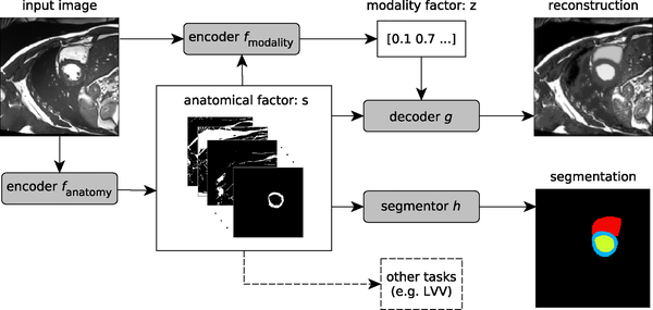
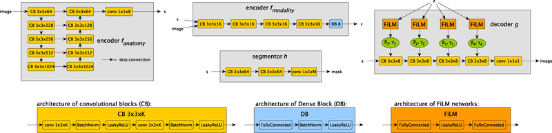
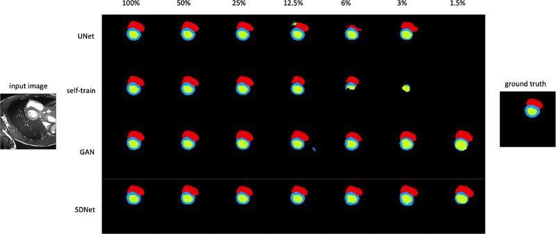
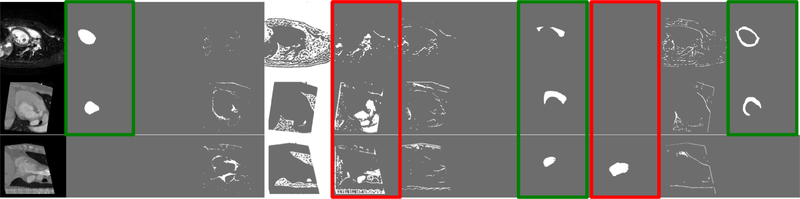
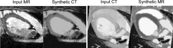
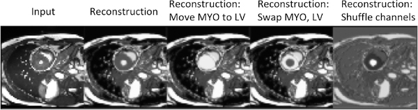
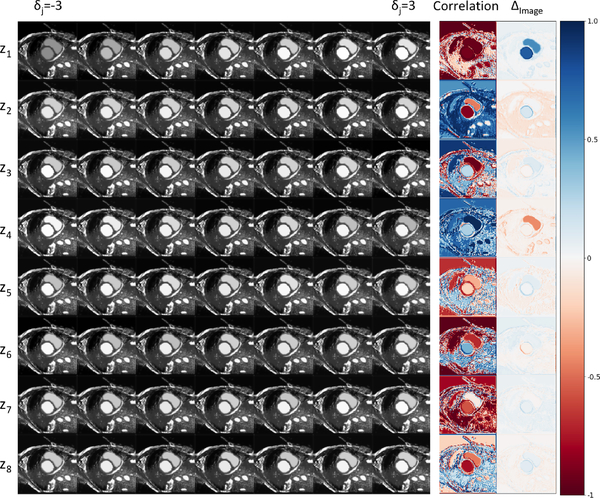
Typically, a medical image offers spatial information on the anatomy (and pathology) modulated by imaging specific characteristics. Many imaging modalities including Magnetic Resonance Imaging (MRI) and Computed Tomography (CT) can be interpreted in this way. We can venture further and consider that a medical image naturally factors into some spatial factors depicting anatomy and factors that denote the imaging characteristics. Here, we explicitly learn this decomposed (disentangled) representation of imaging data, focusing in particular on cardiac images. We propose Spatial Decomposition Network (SDNet), which factorises 2D medical images into spatial anatomical factors and non-spatial modality factors. We demonstrate that this high-level representation is ideally suited for several medical image analysis tasks, such as semi-supervised segmentation, multi-task segmentation and regression, and image-to-image synthesis. Specifically, we show that our model can match the performance of fully supervised segmentation models, using only a fraction of the labelled images. Critically, we show that our factorised representation also benefits from supervision obtained either when we use auxiliary tasks to train the model in a multi-task setting (e.g. regressing to known cardiac indices), or when aggregating multimodal data from different sources (e.g. pooling together MRI and CT data). To explore the properties of the learned factorisation, we perform latent-space arithmetic and show that we can synthesise CT from MR and vice versa, by swapping the modality factors. We also demonstrate that the factor holding image specific information can be used to predict the input modality with high accuracy. Code will be made available at https://github.com/agis85/anatomy_modality_decomposition.
Keywords: Cardiac magnetic resonance imaging; Disentangled representation learning; Multitask learning; Semi-supervised segmentation.
Copyright © 2019. Published by Elsevier B.V.
Figures

References
-
- Almahairi A, Rajeswar S, Sordoni A, Bachman P, Courville AC, 2018. Augmented CycleGAN: Learning many-to-many mappings from unpaired data, in: International Conference on Machine Learning.
-
- Azadi S, Fisher M, Kim V, Wang Z, Shechtman E, Darrell T, 2018. Multi-content GAN for few-shot font style transfer, in: Proceedings of the IEEE Conference on Computer Vision and Pattern Recognition, p. 13.
-
- Bai W, Oktay O, Sinclair M, Suzuki H, Rajchl M, Tarroni G, Glocker B, King A, Matthews PM, Rueckert D, 2017. Semi-supervised learning for network-based cardiac MR image segmentation, in: Medical Image Computing and Computer-Assisted Intervention, Springer International Publishing, Cham: pp. 253–260.
-
- Bai W, Sinclair M, Tarroni G, Oktay O, Rajchl M, Vaillant G, Lee AM, Aung N, Lukaschuk E, Sanghvi MM, Zemrak F, Fung K, Paiva JM, Carapella V, Kim YJ, Suzuki H, Kainz B, Matthews PM, Petersen SE, Piechnik SK, Neubauer S, Glocker B, Rueckert D, 2018a. Automated cardiovascular magnetic resonance image analysis with fully convolutional networks. Journal of Cardiovascular Magnetic Resonance 20, 65. doi: 10.1186/s12968-018-0471-x. - DOI - PMC - PubMed
-
- Bai W, Suzuki H, Qin C, Tarroni G, Oktay O, Matthews PM, Rueckert D, 2018b. Recurrent neural networks for aortic image sequence segmentation with sparse annotations, in: Frangi AF, Schnabel JA, Davatzikos C, Alberola-López C, Fichtinger G (Eds.), Medical Image Computing and Computer Assisted Intervention, Springer International Publishing, Cham: pp. 586–594.
Publication types
MeSH terms
Grants and funding
LinkOut - more resources
Full Text Sources
Other Literature Sources
Medical
