Review
. 2019 Sep;42(9):617-630.
doi: 10.1016/j.tins.2019.06.003. Epub 2019 Jul 24.

Astrocyte-Neuron Interactions in the Striatum: Insights on Identity, Form, and Function

Affiliations
Review

Astrocyte-Neuron Interactions in the Striatum: Insights on Identity, Form, and Function

Baljit S Khakh. Trends Neurosci. 2019 Sep.

Abstract

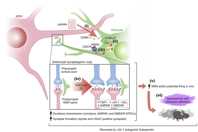
The physiological functions of astrocytes within neural circuits remain incompletely understood. There has been progress in this regard from recent work on striatal astrocytes, where detailed studies are emerging. In this review, findings on striatal astrocyte identity, form, and function, are summarized with a focus on how astrocytes regulate striatal neurons, circuits, and behavior. Specific features of striatal astrocytes are highlighted to illustrate how they may be specialized to regulate medium spiny neurons (MSNs) by responding to, and altering, excitation and inhibition. Further experiments should reveal additional mechanisms for astrocyte-neuron interactions in the striatum and potentially reveal insights into the functions of astrocytes in neural circuits more generally.

Keywords: astrocyte; basal ganglia; behavior; microcircuit; morphology; striatum.


Figures

Figure 1.
Figure 1.. Simplified View of the Striatum in Relation to the Basal Ganglia Circuit and Its Known Roles in Behavior.
(A) Highly schematized cartoon illustrating the striatum in humans in relation to its synaptic inputs [thalamus (T), cortex – associative (A), motor (M), and limbic (L), substantia nigra pars compacta(SNc), and external segment of the globus pallidus(GPe)] and outputs via the basal ganglia (BG) circuitry [GPe, internal segment of the globus pallidus (GPi), and substantia nigra pars reticulate (SNr)]. The striatum receives glutamatergic excitation from multiple regions of the cortex, which impinges on D1 and D2 medium spiny neurons [MSNs (also called striatal projection neurons in some studies); colored red and blue, respectively]. These inputs project to downstream nuclei via the direct and indirect pathway formed by D1 and D2 MSNs, respectively. (B) Cartoon of a tennis player to illustrate that the striatum is intimately involved in movement, action selection, and motor operations. (C) Illustration of the chorea and abnormal movements observed in a patient with advanced-stage Huntington’s disease (HD), whereby movements are uncontrolled and abnormal. Other disorders thought to involve the BG and the striatum are mentioned in the main text. For simplicity, the striatum is shown as a single structure, although in humans it comprises the caudate nucleus, putamen, and nucleus accumbens. Abbreviation: STN, subthalamic nucleus.
Figure 2.
Figure 2.. μ-Crystallin Displays a Gradient of Expression within Striatal Astrocytes.
(A) μ-crystallin immunostaining in striatum showing its spatial gradient. There are higher levels of expression in the ventral striatum compared with dorsal areas. (B) Representative images for μ-crystallin immunostaining in dorsolateral and ventromedial parts of the striatum in brain sections from Aldh1l1-eGFP mice. In the dorsolateral area, ~30% of astrocytes were μ-crystallin positive, whereas, in the ventromedial area, this was ~90%. Adapted from [50]. Abbreviations: Cc, corpus callosum; Ctx, cortex; V, ventricle.
Figure 3.
Figure 3.. Properties of Striatal Astrocytes.
(A) Coronal sections of Aldh1l1-eGFP mouse brains cleared using the Sca/eS method and imaged using confocal microscopy to show abundant astrocytes within the striatum. The magnified section on the right shows the dorsolateral region of the striatum. Blood vessels are demarcated by their associations with GFP-expressing astrocyte end-feet. (B) Confocal volumes of a Lucifer yellow-filled striatal astrocyte. (C) 3D reconstructions of volumes enclosed by striatal astrocyte territories (blue) and NeuN (red). (D) Example of scanning electron microscopy (SEM) image from the striatum with corresponding 3D rendering displayed at an angle. The synaptic structures and closest astrocyte processes are colored as follows: yellow, astrocytes; blue, postsynaptic densities (PSDs); green, axons; and pink, spines. The center of the PSD is denoted by a red dot. (E) Representative current waveforms for a striatal astrocyte in response to stepwise changes membrane potential, along with an average current voltage relation from multiple cells. Reproduced from [50] (A–D); data in (E) from [51].
Figure 4.
Figure 4.. Working Model for How Attenuating Striatal Astrocyte Ca2+-Dependent Signaling In Vivo Altered Striatal Neural Circuit Function with Behavioral Consequences.
The cartoon summary is of the main findings at synaptic (A) and in vivo levels (B). (C) Description of the proposed sequence of events. In brief, attenuation of striatal astrocyte Ca2+ signals reduces Rab11a, which results in increased GAT-3 functional expression. This reduces ambient γ-aminobutyric acid (GABA) levels in the extracellular space and tonic inhibition. The data are consistent with a model in which reduced tonic inhibition alters medium spiny neurons (MSN) firing and downstream circuits to cause excessive self-grooming. In accord, tonic inhibition and self-grooming were rescued by a GAT-3 antagonist. Reproduced from [52].
Figure 5.
Figure 5.. A Schematized Working Model for how Gi G-Protein-Coupled Receptor (GPCR)-Mediated Medium Spiny Neuron (MSN)–Astrocyte Bidirectional Interactions Affect Behavior.
When MSNs were depolarized to levels associated with upstates, they released γ-aminobutyric acid (GABA) (step i), which activated Gi-protein coupled GABAB G-protein-coupled receptors (GPCRs) on striatal astrocytes, leading to an increase in intracellular Ca2+ signals (step ii). Selectively stimulating the Gi pathway with hM4Di and clozapine-n-oxide (CNO) evoked Ca2+ signals in striatal astrocytes (step iii), upregulated the astrocyte synaptogenic molecule TSP-1, boosted excitatory synapse formation and fast excitatory synaptic transmission (step iv) and increased firing of MSNs (step v), which together resulted in hyperactivity with disrupted attention phenotypes in mice (step vi). The synaptic, circuit, and behavioral effects resulting from Gi pathway activation in vivo (steps iv–vi) were reduced or reversed by blocking TSP-1 actions on neuronal α2δ–1 receptors with gabapentin. Reproduced from [53]. Abbreviation: EPSC, excitatory postsynaptic current.

References

    1. Kettenmann H and Verkhratsky A (2008) Neuroglia: the 150 years after. Trends Neurosci. 31,653–659 - PubMed
    1. Allen NJ and Lyons DA (2018) Glia as architects of central nervous system formation and function. Science 362, 181–185 - PMC - PubMed
    1. Shepherd GM and Grillner S (2010) Handbook of Brain Microcircuits, Oxford University Press
    1. Khakh BS and Sofroniew MV (2015) Diversity of astrocyte functions and phenotypes in neural circuits. Nat. Neurosci 18, 942–952 - PMC - PubMed
    1. Erö C et al. (2018) A cell atlas for the mouse brain. Front. Neuroinform 12, 84. - PMC - PubMed

Publication types