. 2019 Nov:63:109366.
doi: 10.1016/j.cellsig.2019.109366. Epub 2019 Jul 25.

Arrestin-3 interaction with maternal embryonic leucine-zipper kinase

Affiliations

Arrestin-3 interaction with maternal embryonic leucine-zipper kinase

Nicole A Perry et al. Cell Signal. 2019 Nov.

Abstract

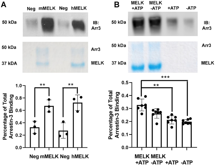
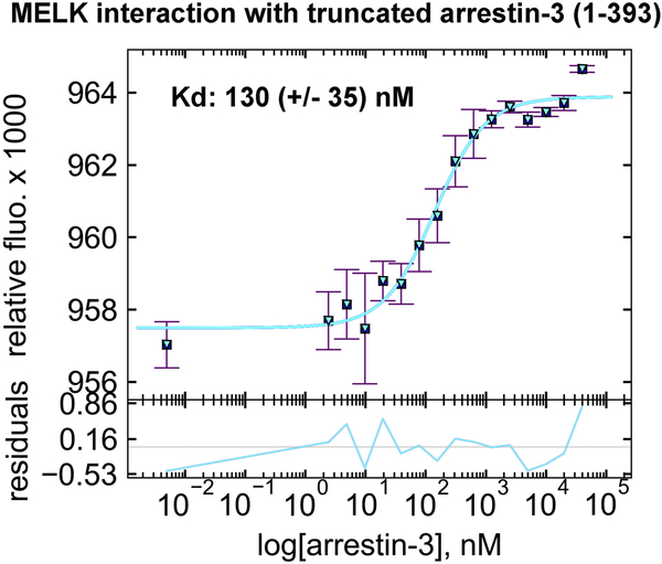
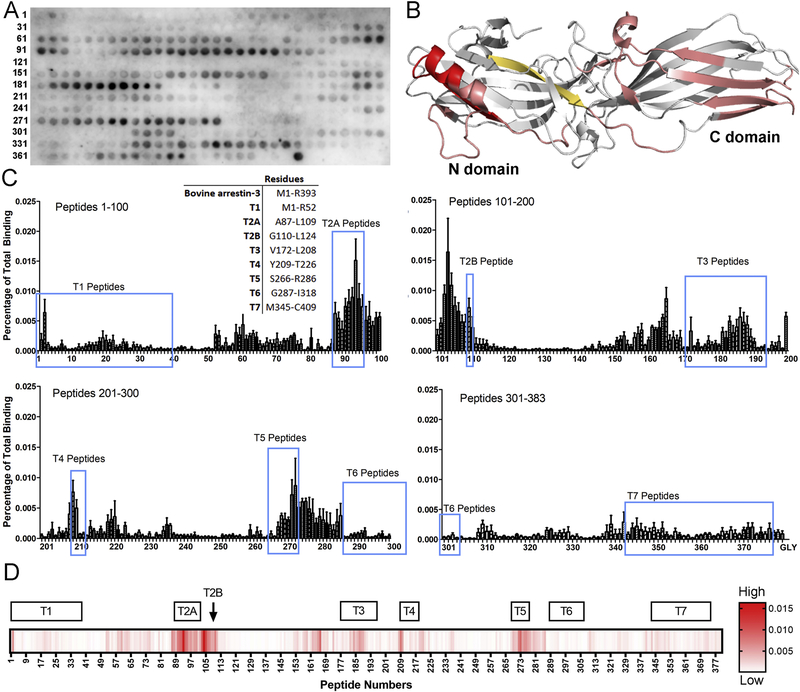
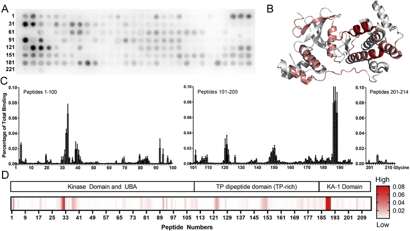
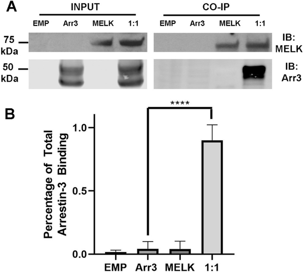
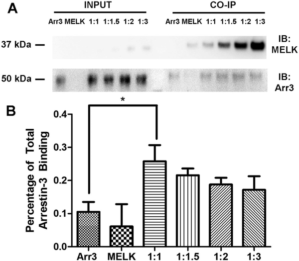
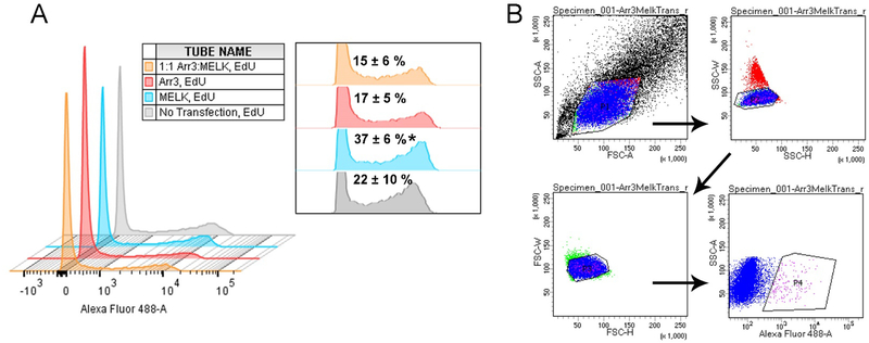
Maternal embryonic leucine-zipper kinase (MELK) overexpression impacts survival and proliferation of multiple cancer types, most notably glioblastomas and breast cancer. This makes MELK an attractive molecular target for cancer therapy. Yet the molecular mechanisms underlying the involvement of MELK in tumorigenic processes are unknown. MELK participates in numerous protein-protein interactions that affect cell cycle, proliferation, apoptosis, and embryonic development. Here we used both in vitro and in-cell assays to identify a direct interaction between MELK and arrestin-3. A part of this interaction involves the MELK kinase domain, and we further show that the interaction between the MELK kinase domain and arrestin-3 decreases the number of cells in S-phase, as compared to cells expressing the MELK kinase domain alone. Thus, we describe a new mechanism of regulation of MELK function, which may contribute to the control of cell fate.

Keywords: Arrestin; Cell fate signaling; Maternal embryonic leucine-zipper kinase (MELK); Protein-protein interactions.


Conflict of interest statement

Conflict of Interest

The authors declare no conflict of interest.

Figures

Figure 1.
Figure 1.. Pull-down of purified MELK1−326,T167E with purified arrestin-31–393.
(A) Analysis of mouse MELK1−326,T167E (10 μg; mMELK) and human MELK1−326,T167E (10 μg; hMELK) binding to bovine arrestin-31−393 (10 μg). Top: Representative Western blot of the eluates using a polyclonal anti-arrestin antibody (1:10,000, F431 [58]). Middle: Representative Coomassie staining of the eluates. Bottom: Quantification of arrestin-3 binding using densitometry. Intensity was compared to the negative control (beads alone) and binding is shown as a percentage of total arrestin-3 applied (n=3). Statistical analysis was performed using Student’s t-test (**, p<0.01). (B) Analysis of human MELK1−326,T167E (10 μg) binding to bovine arrestin-31−393 (10 μg) in the absence or presence of 2 mM ATP and 4 mM MgCl2. Top: Representative Western blot of the eluates using an anti-arrestin-3 antibody (1:10,000, F431). Middle: Representative Coomassie staining of the eluates. Bottom: Quantification of arrestin-3 binding using densitometry. Intensity was compared to the negative control (beads alone) and binding is displayed as a percentage of total arrestin-3 applied (n=7). Statistical analysis was performed using Kruskal-Wallis test with multiple comparisons (**, p<0.01; ***, p<0.001).
Figure 2.
Figure 2.. The affinity of MELK1−326,T167E for arrestin-31−393 in the presence of ATP.
A binding curve for bovine arrestin-31−393 with human MELK1−326,T167E in the presence of 1 mM ATP and 2 mM MgCl2 is shown with the average Kd value (n=3). Microscale thermophoresis was performed at a constant concentration of Tris-NTA-labeled His-MELK1−326,T167E (50 nM) with a serial dilution of arrestin-31−393 (0–40 μM). The binding isotherm was calculated using preset T-jump in the software PALMIST [59, 60], and the graph was created in the program GUSSI.
Figure 3.
Figure 3.. MELK1−326,T167E binding sites on arrestin-3.
(A) Representative peptide blot (15-mer) of bovine arrestin-3-derived peptides (residues 1–393, 1 amino acid shifts). (B) Map of interaction sites for MELK1−326,T167E on arrestin-3 (PDB 3P2D [40]). Peptides calculated to be in the top 1% for binding to MELK1−326,T167E are highlighted in red and top 10% in salmon. The peptide V165-Q173 (shown in orange-yellow) demonstrated binding within the top 10% but is likely a false positive due to its surface inaccessibility in both basal [40] and active [33] arrestin-3 structures. (C) Quantified binding of MELK1−326,T167E to arrestin-3-derived peptides. Binding was measured as a percentage of total intensity on the membrane. A 15-mer glycine peptide was used to determine non-specific interactions. Inset: Peptide segments corresponding to the non-receptor surface of arrestin-3. The corresponding regions are circled in blue on the quantification. (D) Heat map using a single gradient to display ranges of binding was calculated using GraphPad Prism 8.0.2. Peptides corresponding to the non-receptor-binding surface of arrestin-3 are shown above.
Figure 4.
Figure 4.. Arrestin-31−393 binding sites on MELK1–651.
(A) Representative peptide blot using 15-mers derived from human MELK (residues 1–651, 3 amino acid shifts). (B) Map of interaction sites for arrestin-31−393 on MELK (PDB 4BL1; to be published [45]). Peptides were analyzed using conditional formatting and those that exhibited binding to arrestin-31−393 within the top 10% are highlighted in salmon, while those binding within the top 5% are shown in red. The crystal structure of the MELK kinase domain used a construct that is truncated after residue 347 (peptide 107 in the array). Therefore, the high binding peptides from the C-terminal KA1 domain (188–190; sequence NVTTTRLVNPDQLLNEIMSIL), cannot be mapped onto the crystal structure. (C) Quantified binding of arrestin-31−393 to MELK-derived peptides. Arrestin-31−393 binding was measured as a percentage of total intensity of all spots. (D) Heat map using a single gradient to display ranges of binding was calculated using GraphPad Prism 8.0.2. Corresponding MELK domains are shown above.
Figure 5.
Figure 5.. Co-immunoprecipitation of wild-type MELK1−651 with arrestin-31–393.
(A) Western analysis of coimmunoprecipitation. HEK293 arrestin-2/3 knockout cells were transfected with empty vector (20 μg), HA-arrestin-31−393 (10 μg), FLAG-MELK1−651 (10 μg), or both plasmids encoding arrestin-3 and MELK at a 1:1 DNA ratio for 48 h prior to immunoprecipitation with FLAG primary antibody. Western analysis was performed using anti-arrestin (1:10,000; F431) and anti-FLAG (1: 1,000; F3165 Sigma) antibodies. (B) Arrestin-31−393 co-immunoprecipitation with MELK1−651 was quantified using densitometric analysis of arrestin-3 blots. Empty vector and non-specific arrestin-3 binding (no bait control) are also shown. Statistical analysis was performed using One-way ANOVA followed by Dunnett’s post hoc test with correction for multiple comparisons (****, p<0.0001).
Figure 6.
Figure 6.. Co-immunoprecipitation of wild-type MELK1−340 with arrestin-31–393.
(A) Western analysis of coimmunoprecipitation. HEK293 arrestin-2/3 knockout cells were transfected with HA-arrestin-31−393 (1 μg), FLAG-MELK1−340 (1 μg), or both plasmids encoding arrestin-3 and MELK at indicated DNA ratios for 48 h prior to immunoprecipitation with FLAG primary antibody. Western analysis was performed using anti-arrestin (1:10,000; F431) and anti-FLAG (1: 1,000; F3165 Sigma) antibodies. (B) Arrestin-31−393 co-immunoprecipitation with MELK1−340 was quantified using densitometric analysis of arrestin-3 blots. Non-specific arrestin-3 binding (no bait control) is also shown. Statistical analysis was performed using One-way ANOVA followed by Tukey’s post hoc test with correction for multiple comparisons (*, p<0.05).
Figure 7.
Figure 7.. The effect of wild-type MELK1−340 and arrestin-31−393 expression on the fraction of cells in S-phase.
HEK293 arrestin-2/3 knockout cells were transfected with HA-arrestin-31−393 (1 μg) (red), FLAG-MELK1−340 (1 μg) (blue), or a fixed DNA ratio of plasmids encoding the two proteins (1 μg:1 μg) (orange), and the number of cells in S-phase was measured using the Click-iT™ EdU Alexa Fluor 488 Imaging Kit (ThermoFisher Scientific), followed by flow cytometry. (A) A representative histogram shows data from cells labeled with Alexa Fluor 488 picolyl azide analyzed using a 3-laser LSRII (BD Biosciences). The inset shows the relative heights of the second distribution (Alexa Fluor 488 positive cells) with the percentages of cells in S-phase for combined experiments (n=3). Analysis was completed on FlowJo 10.1.5 (FlowJo LLC). Statistical analysis was performed using One-way ANOVA followed by Tukey’s post-hoc test (*, p<0.05 to 1:1 Arr3:MELK). The first distribution represents cells that are not proliferating, while the second distribution represents cells in S-phase. (B) The gating strategy used for histogram generation. Cells were initially sorted by scatter (SSC-A by FSC-A) (P1) to determine the population of living cells, then by single cells (SSC-W by SSC-H) (P2), then singlets (FSC-W by FSC-H) (P3), and finally by cells positive for Alexa Fluor 488 (SSC-A by Alexa Fluor 488) (P4).

Similar articles

Cited by

References

    1. Ganguly R, Mohyeldin A, Thiel J, Kornblum HI, Beullens M, Nakano I. MELK—a conserved kinase: functions, signaling, cancer, and controversy. Clinical and Translational Medicine. 2015;4:11. - PMC - PubMed
    1. Lizcano JM, Göransson O, Toth R, Deak M, Morrice NA, Boudeau J, et al. LKB1 is a master kinase that activates 13 kinases of the AMPK subfamily, including MARK/PAR-1. EMBO J. 2004;23:833–43. - PMC - PubMed
    1. Jung H, Seong H-A, Ha H. Murine Protein Serine/Threonine Kinase 38 Activates Apoptosis Signal-regulating Kinase 1 via Thr838 Phosphorylation. Journal of Biological Chemistry. 2008;283:34541–53. - PMC - PubMed
    1. Giuliano CJ, Lin A, Smith JC, Palladino AC, Sheltzer JM. MELK expression correlates with tumor mitotic activity but is not required for cancer growth. eLife. 2018;7:e32838. - PMC - PubMed
    1. Speers C, Zhao SG, Kothari V, Santola A, Liu M, Wilder-Romans K, et al. Maternal Embryonic Leucine Zipper Kinase (MELK) as a Novel Mediator and Biomarker of Radioresistance in Human Breast Cancer. Clinical Cancer Research. 2016;22:5864. - PMC - PubMed

Publication types

LinkOut - more resources