. 2019 Sep 5;105(3):631-639.
doi: 10.1016/j.ajhg.2019.07.002. Epub 2019 Jul 25.

Haploinsufficiency of the Notch Ligand DLL1 Causes Variable Neurodevelopmental Disorders

Affiliations

Haploinsufficiency of the Notch Ligand DLL1 Causes Variable Neurodevelopmental Disorders

Björn Fischer-Zirnsak et al. Am J Hum Genet. .

Abstract

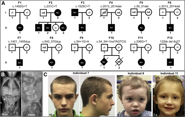
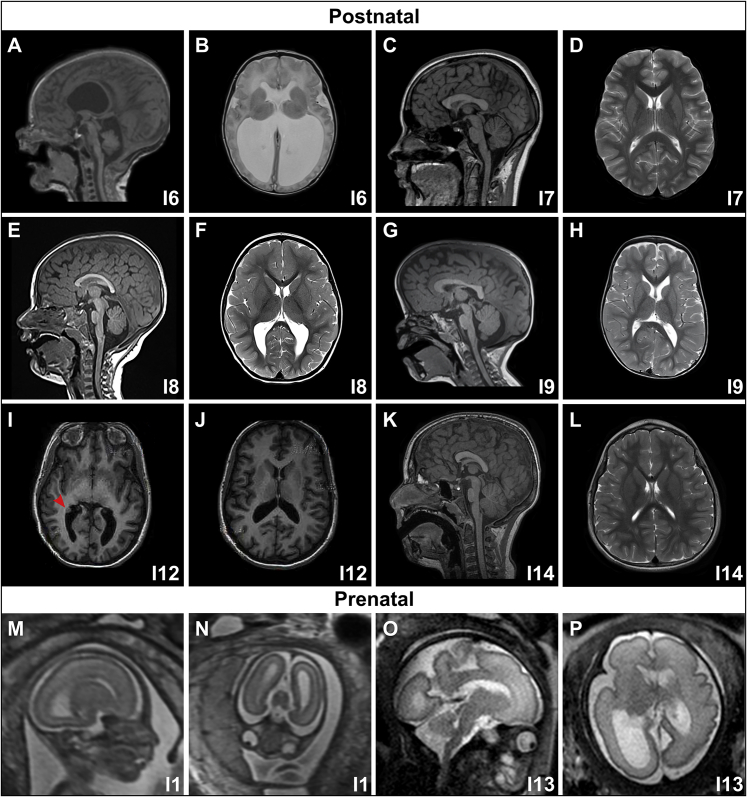
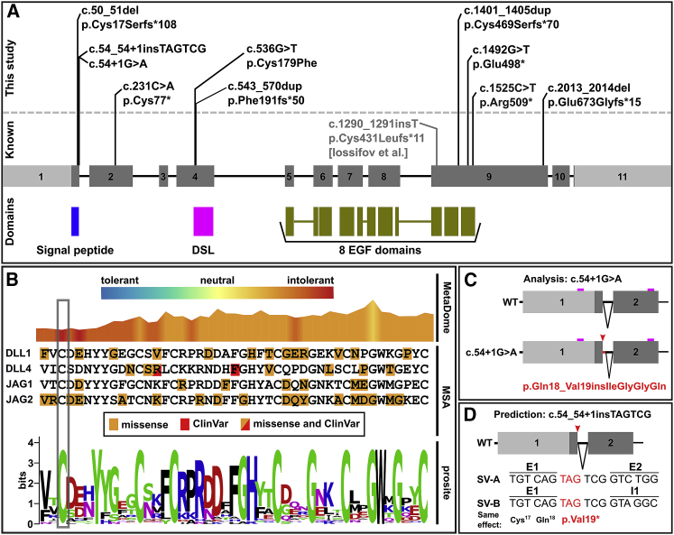
Notch signaling is an established developmental pathway for brain morphogenesis. Given that Delta-like 1 (DLL1) is a ligand for the Notch receptor and that a few individuals with developmental delay, intellectual disability, and brain malformations have microdeletions encompassing DLL1, we hypothesized that insufficiency of DLL1 causes a human neurodevelopmental disorder. We performed exome sequencing in individuals with neurodevelopmental disorders. The cohort was identified using known Matchmaker Exchange nodes such as GeneMatcher. This method identified 15 individuals from 12 unrelated families with heterozygous pathogenic DLL1 variants (nonsense, missense, splice site, and one whole gene deletion). The most common features in our cohort were intellectual disability, autism spectrum disorder, seizures, variable brain malformations, muscular hypotonia, and scoliosis. We did not identify an obvious genotype-phenotype correlation. Analysis of one splice site variant showed an in-frame insertion of 12 bp. In conclusion, heterozygous DLL1 pathogenic variants cause a variable neurodevelopmental phenotype and multi-systemic features. The clinical and molecular data support haploinsufficiency as a mechanism for the pathogenesis of this DLL1-related disorder and affirm the importance of DLL1 in human brain development.

Keywords: DLL1; Notch signaling; autism; brain malformation; developmental delay; intellectual disability; vertebral segmentation defects.


Conflict of interest statement

E.T., A.T., Y.S., Y.C., S.L., K.M., and X.W. are employees of GeneDx, Inc., a wholly owned subsidiary of OPKO Health, Inc. All other authors declare no competing interests.

Figures

Figure 1
Figure 1
Pedigrees of the Affected Individuals and Phenotypic Presentation (A) Twelve unrelated families with DLL1 pathogenic variants. The DLL1 variants occurred de novo in families (F) 1, 6, 7, 9, 10, 11, and 12. The DLL1 variant co-segregated with the phenotype in F2 and F3. Inheritance is unknown due to lack of parenteral DNA samples for F4, F5, and F8. (B) Radiographs of individual (I) 8 (F5/II-1 in A) at the age of 10 months demonstrate lumbar scoliosis (34°) due to a segmentation defect of the lumbar spine; the vertebral malformations included an incomplete fusion of the vertebral arch in L1, a right L2 hemivertebra, an abnormally shaped L3, and an asymmetric S1. (C) Facial photographs of I7 (F4/II-1), I9 (F6/II-1), and I11 (F8/II-1) showing upslanted palpebral fissures and mild retrognathia (I7), a prominent forehead, upslanted palpebral fissures, epicanthal folds, broad and flat nasal bridge, full cheeks, everted upper lip, and full lips (I9) as well as upslanted palpebral fissures (I11).
Figure 2
Figure 2
Representative Brain MR Images of the DLL1 Cohort (A and B) T1-midsagittal (A) and T2-axial (B) images of I6 showing severe hydrocephalus with a thin/stretched corpus callosum, thin brainstem, small cerebellum, and compression of the overlying cortex. (C and D) T1-midsagittal (C) and T2-axial (D) images of I7 showing a mildly short and thick corpus callosum, and subtle areas of cortical dysplasia. (E and F) T1-midsagittal (E) and T2-axial (F) images of I8 showing a mildly dysplastic corpus callosum, mild cortical dysplasia, and mild ventriculomegaly. (G and H) T1-midsagittal (G) and T2-axial (H) images of I9 showing relatively large brain size with a prominent forehead, mildly dysplastic corpus callosum, and mildly dilated ventricles. (I and J) T1-axial images of I12 showing PVNH (red arrowhead, I) with mild ventriculomegaly. (K and L) T1-midsagittal (K) and T2-axial (L) images of I14 showing a mildly short and dysplastic corpus callosum, mildly small pons, and subtle cortical dysplasia. (M and N) Prenatal T2 sagittal (M) and axial (N) images of I1 showing ventriculomegaly, with no good mid-sagittal views. (O and P) Prenatal T2 sagittal (O) and axial (P) images of I13 showing asymmetric ventriculomegaly.
Figure 3
Figure 3
Spectrum of DLL1 Pathogenic Variants and Functional Interpretations (A) Schematic of the DLL1 gene with DLL1 variants annotated. A previously described LoF variant is indicated in gray. The DLL1 protein domains are listed below. (B) Sequence and variant characteristics of the DSL domain. The missense variant GenBank: NM_005618.3; p.Cys179Phe of I14 is highlighted in gray. Top: visualization of the MetaDome score. Center: multi sequence alignment (MSA) of the DSL domain of genes annotated with DSL domain by Prosite (DLL1, DLL4, JAG1, JAG4). Positions with missense variants in gnomAD version 2.1 as well as pathogenic ClinVar variants are labeled (no frameshift and nonsense variants are present in gnomAD). Bottom: Conservation of the DSL domain (Prosite entry PS51051). Cysteine is conserved among DSL domains. The MetaDome score indicates the deleterious nature of any amino acid changes at this position. (C) Scheme of the consequence of the splice site variant identified in I12. Alternative splicing of the allele carrying c.54+1G>A leads to the retention of 12 intronic base pairs (bp), resulting in an in-frame insertion of four amino acids (p.Gln18_Val19insIleGlyGlyGln). (D) Predicted consequence of the insertion c.54_54+1insTAGTCG identified in I13. Human Splicing Finder predicted that the insertion c.54_54+1insTAGTCG leads to an alternative donor splice site after the insertion, resulting in two different predicted transcript forms. Both lead to a premature stop codon. Boxes present exons, lines present introns; light gray boxes: untranslated regions; dark gray boxes: coding sequence; small boxes in magenta in (C): location of the sequencing primers; red arrows: location of the splice variants; black triangles: intronic sequence removed by splicing; SV-A: splice variant A; SV-B: splice variant B. E1 and E2 represent sequence from exon 1 and 2 respectively, I1 represents intron 1.

References

    1. Bray S.J. Notch signalling in context. Nat. Rev. Mol. Cell Biol. 2016;17:722–735. - PubMed
    1. Kakuda S., Haltiwanger R.S. Deciphering the Fringe-Mediated Notch Code: Identification of Activating and Inhibiting Sites Allowing Discrimination between Ligands. Dev. Cell. 2017;40:193–201. - PMC - PubMed
    1. Louvi A., Artavanis-Tsakonas S. Notch signalling in vertebrate neural development. Nat. Rev. Neurosci. 2006;7:93–102. - PubMed
    1. Bettenhausen B., Hrabĕ de Angelis M., Simon D., Guénet J.L., Gossler A. Transient and restricted expression during mouse embryogenesis of Dll1, a murine gene closely related to Drosophila Delta. Development. 1995;121:2407–2418. - PubMed
    1. Hrabĕ de Angelis M., McIntyre J., 2nd, Gossler A. Maintenance of somite borders in mice requires the Delta homologue DII1. Nature. 1997;386:717–721. - PubMed

Publication types