

Delayed delivery of endothelial progenitor cell-derived extracellular vesicles via shear thinning gel improves postinfarct hemodynamics
- PMID: 31353103
- PMCID: PMC7077034
- DOI: 10.1016/j.jtcvs.2019.06.017
Delayed delivery of endothelial progenitor cell-derived extracellular vesicles via shear thinning gel improves postinfarct hemodynamics
Abstract
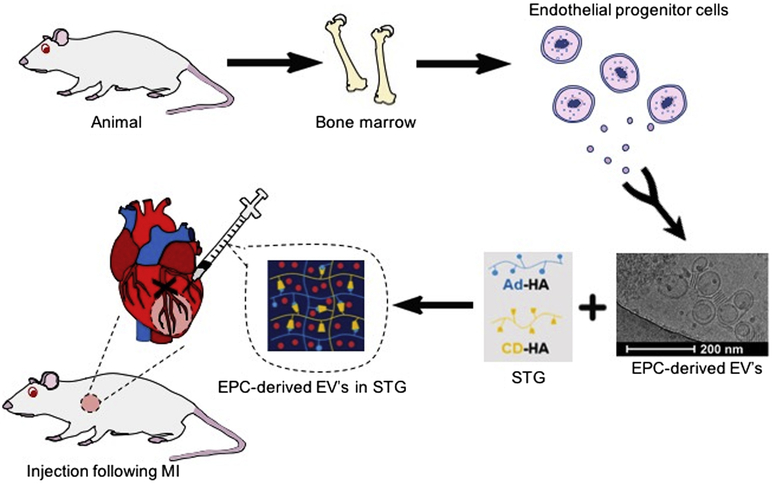
Background: Extracellular vesicles (EVs) are promising therapeutics for cardiovascular disease, but poorly-timed delivery might hinder efficacy. We characterized the time-dependent response to endothelial progenitor cell (EPC)-EVs within an injectable shear-thinning hydrogel (STG+EV) post-myocardial infarction (MI) to identify when an optimal response is achieved.
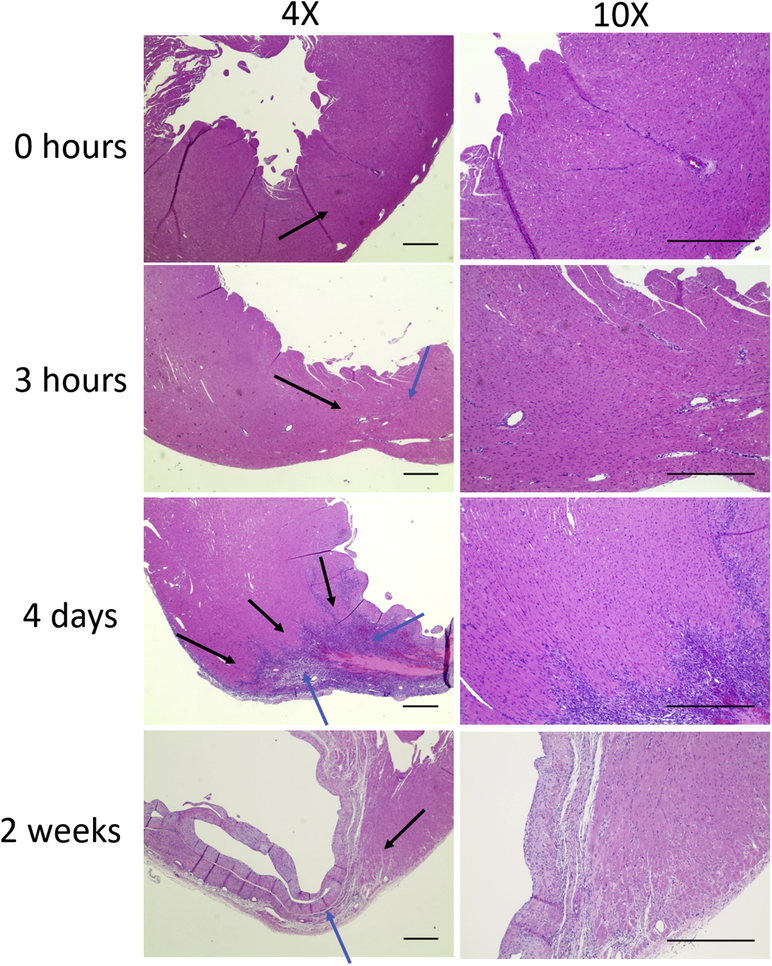
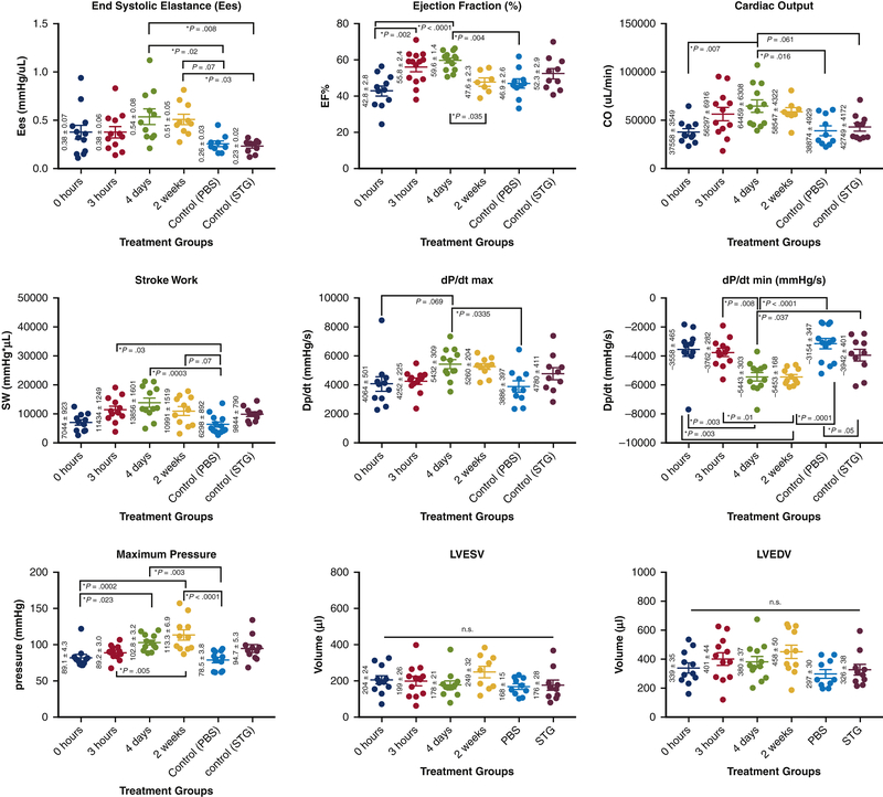
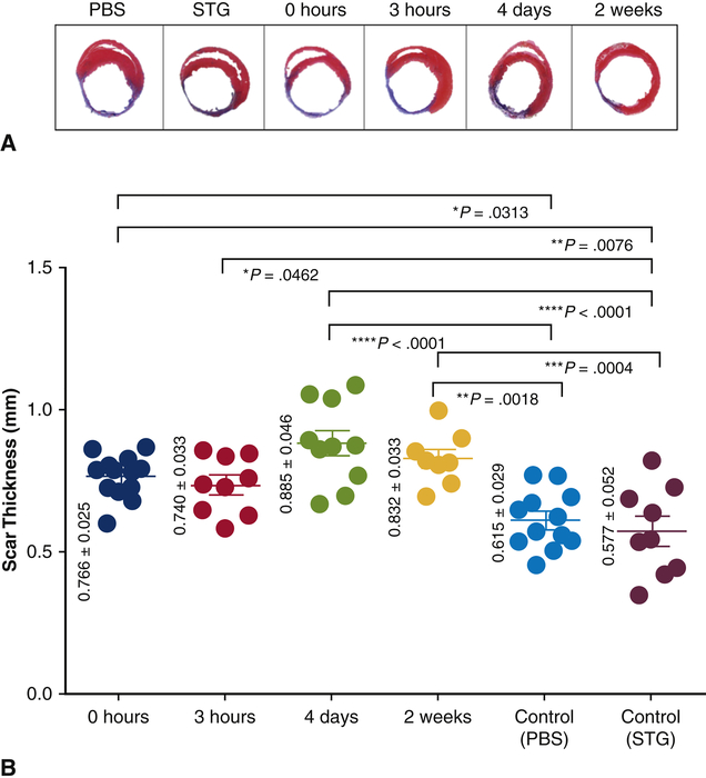
Methods: The angiogenic effects of prolonged hypoxia on cell response to EPC-EV therapy and EV uptake affinity were tested in vitro. A rat model of acute MI via left anterior descending artery ligation was created and STG+EV was delivered via intramyocardial injections into the infarct border zone at time points corresponding to phases of post-MI inflammation: 0 hours (immediate), 3 hours (acute inflammation), 4 days (proliferative), and 2 weeks (fibrosis). Hemodynamics 4 weeks post-treatment were compared across treatment and control groups (phosphate buffered saline [PBS], shear-thinning gel). Scar thickness and ventricular diameter were assessed histologically. The primary hemodynamic end point was end systolic elastance. The secondary end point was scar thickness.
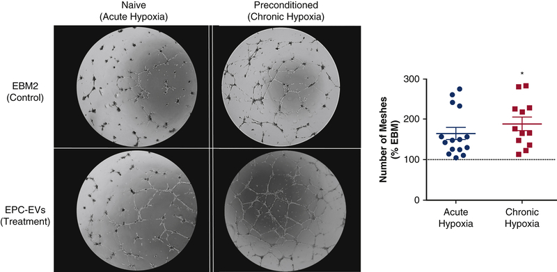
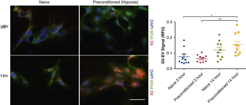
Results: EPC-EVs incubated with chronically versus acutely hypoxic human umbilical vein endothelial cells resulted in a 2.56 ± 0.53 versus 1.65 ± 0.15-fold increase (P = .05) in a number of vascular meshes and higher uptake of EVs over 14 hours. End systolic elastance improved with STG+EV therapy at 4 days (0.54 ± 0.08) versus PBS or shear-thinning gel (0.26 ± 0.03 [P = .02]; 0.23 ± 0.02 [P = .01]). Preservation of ventricular diameter (6.20 ± 0.73 mm vs 8.58 ± 0.38 mm [P = .04]; 9.13 ± 0.25 mm [P = .01]) and scar thickness (0.89 ± 0.05 mm vs 0.62 ± 0.03 mm [P < .0001] and 0.58 ± 0.05 mm [P < .0001]) was significantly greater at 4 days, compared wit PBS and shear-thinning gel controls.
Conclusions: Delivery of STG+EV 4 days post-MI improved left ventricular contractility and preserved global ventricular geometry, compared with controls and immediate therapy post-MI. These findings suggest other cell-derived therapies can be optimized by strategic timing of therapeutic intervention.
Keywords: delayed therapy; extracellular vesicles; myocardial infarction; shear thinning gel.
Copyright © 2019 The American Association for Thoracic Surgery. Published by Elsevier Inc. All rights reserved.
Conflict of interest statement
Disclosure: None of the authors have any conflicts of interest
Figures
Comment in
-
Commentary: "Shear" patience for post-myocardial infarction regenerative therapy.J Thorac Cardiovasc Surg. 2020 May;159(5):1836-1837. doi: 10.1016/j.jtcvs.2019.06.033. Epub 2019 Jul 2. J Thorac Cardiovasc Surg. 2020. PMID: 31327546 Free PMC article. No abstract available.
-
Commentary: All things have their season-Timing of regenerative treatment.J Thorac Cardiovasc Surg. 2020 May;159(5):1838-1839. doi: 10.1016/j.jtcvs.2019.06.069. Epub 2019 Jul 8. J Thorac Cardiovasc Surg. 2020. PMID: 31371107 No abstract available.
References
Publication types
MeSH terms
Substances
Grants and funding
LinkOut - more resources
Full Text Sources
Medical
