. 2019 Aug 19;29(16):2625-2639.e5.
doi: 10.1016/j.cub.2019.06.062. Epub 2019 Jul 25.

Structure of the Human Core Centromeric Nucleosome Complex

Affiliations

Structure of the Human Core Centromeric Nucleosome Complex

Praveen Kumar Allu et al. Curr Biol. .

Abstract

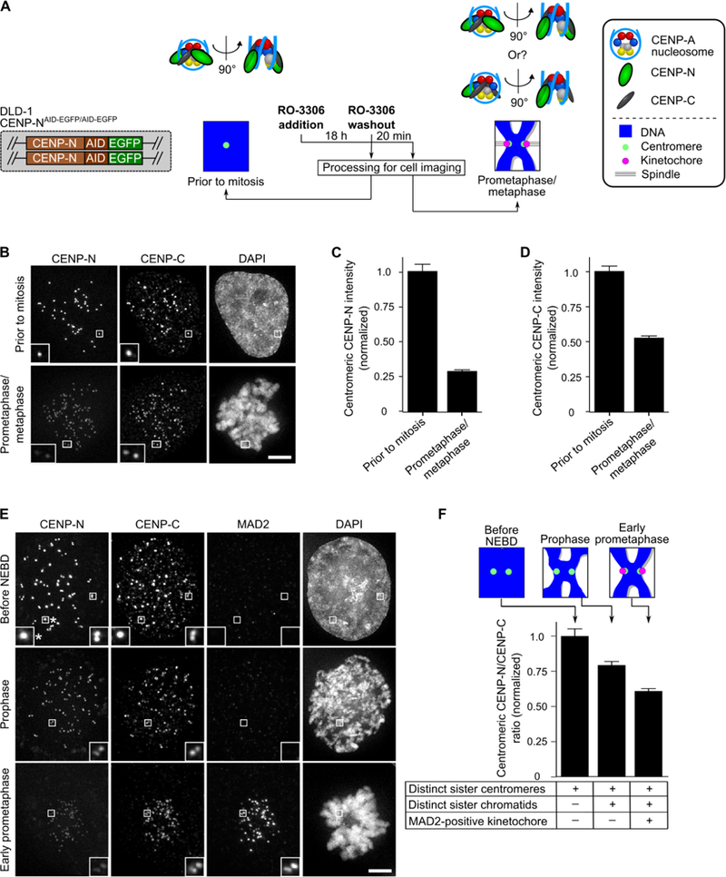
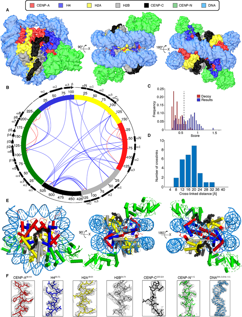
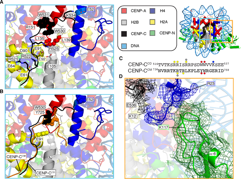
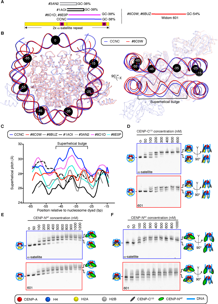
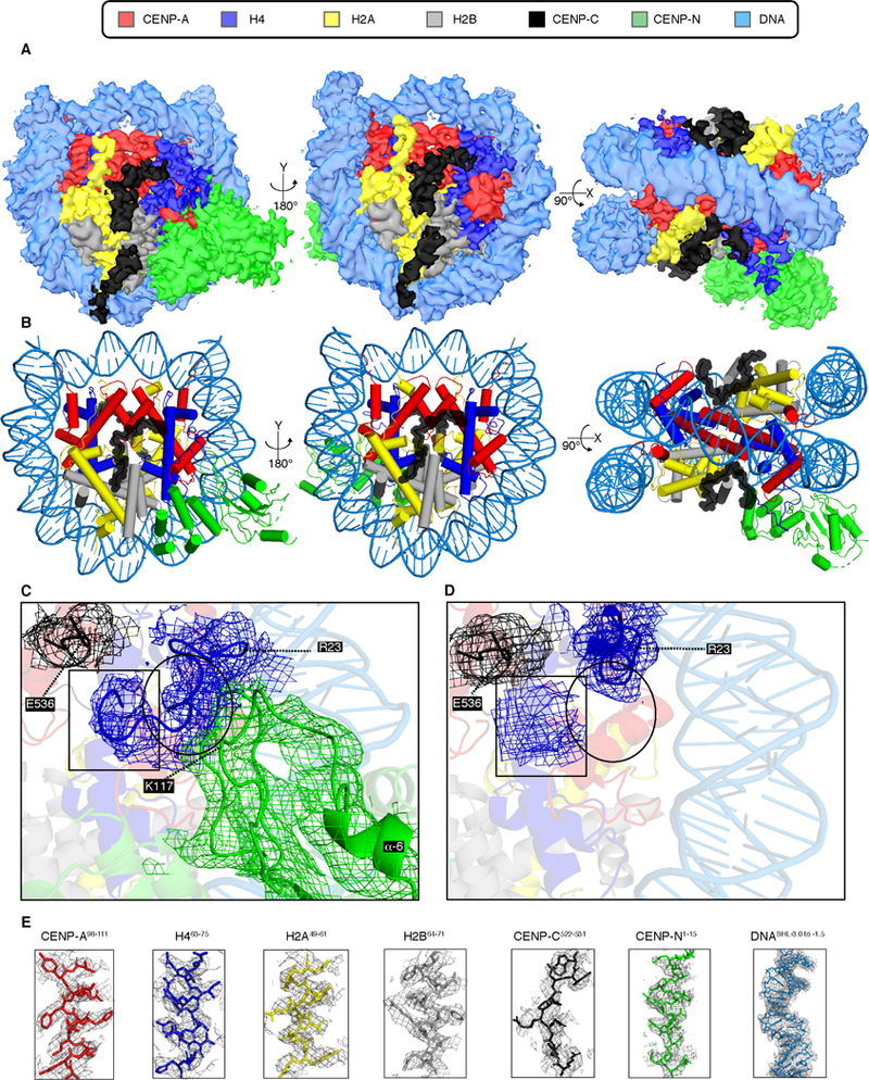
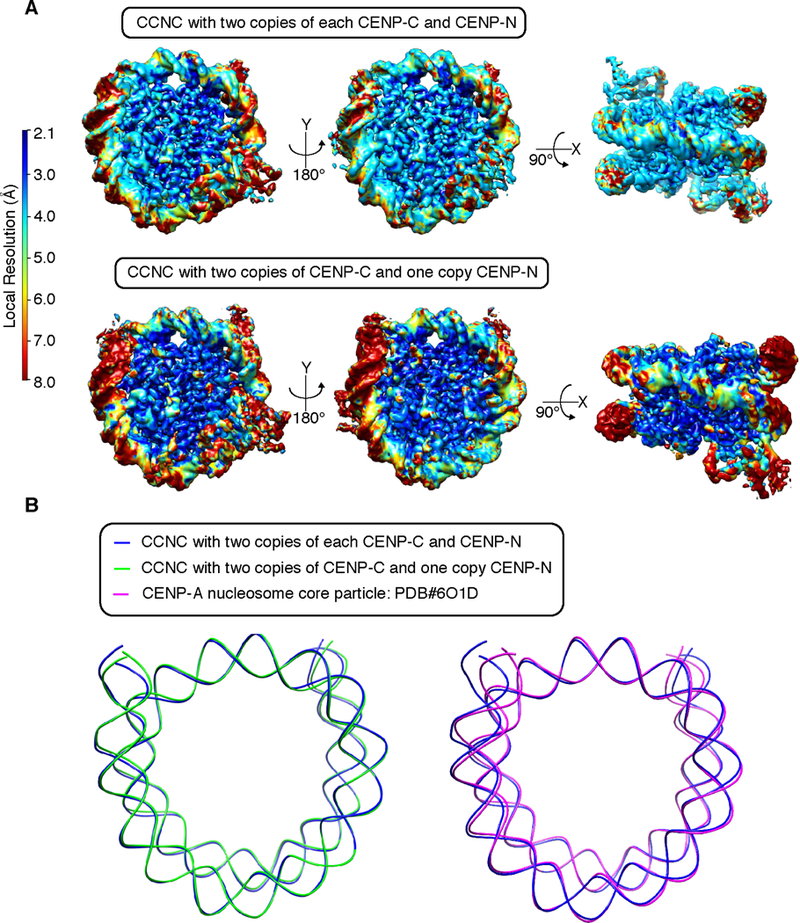
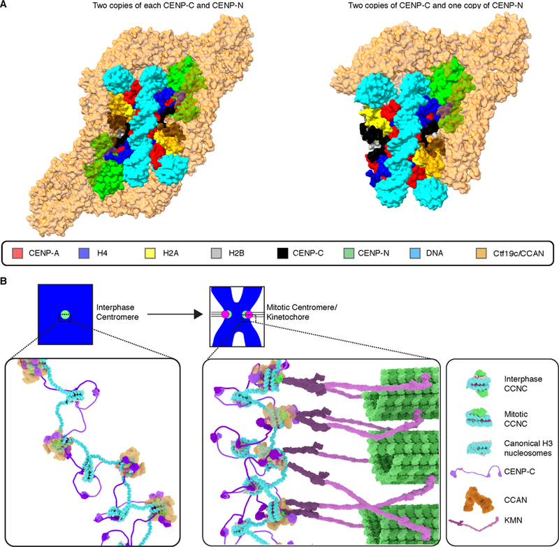
Centromeric nucleosomes are at the interface of the chromosome and the kinetochore that connects to spindle microtubules in mitosis. The core centromeric nucleosome complex (CCNC) harbors the histone H3 variant, CENP-A, and its binding proteins, CENP-C (through its central domain; CD) and CENP-N (through its N-terminal domain; NT). CENP-C can engage nucleosomes through two domains: the CD and the CENP-C motif (CM). CENP-CCD is part of the CCNC by virtue of its high specificity for CENP-A nucleosomes and ability to stabilize CENP-A at the centromere. CENP-CCM is thought to engage a neighboring nucleosome, either one containing conventional H3 or CENP-A, and a crystal structure of a nucleosome complex containing two copies of CENP-CCM was reported. Recent structures containing a single copy of CENP-NNT bound to the CENP-A nucleosome in the absence of CENP-C were reported. Here, we find that one copy of CENP-N is lost for every two copies of CENP-C on centromeric chromatin just prior to kinetochore formation. We present the structures of symmetric and asymmetric forms of the CCNC that vary in CENP-N stoichiometry. Our structures explain how the central domain of CENP-C achieves its high specificity for CENP-A nucleosomes and how CENP-C and CENP-N sandwich the histone H4 tail. The natural centromeric DNA path in our structures corresponds to symmetric surfaces for CCNC assembly, deviating from what is observed in prior structures using artificial sequences. At mitosis, we propose that CCNC asymmetry accommodates its asymmetric connections at the chromosome/kinetochore interface. VIDEO ABSTRACT.

Keywords: cell division; centromere; chromatin; cryo-EM; epigenetics; histone; kinetochore; microtubule spindle; mitosis; nucleosome.


Conflict of interest statement

Declaration of interest

The authors declare no conflict of interest.

Figures

Figure 1.
Figure 1.. CENP-C is quantitatively retained at centromeres upon mitotic entry, but CENP-N levels drop by half prior to kinetochore formation.
(A) Schematic representation for measuring CCNC components upon mitotic entry. (B) Representative images of cells treated as in panel A. Scale bar, 5 µm. (C,D) Quantification of experiment in panel B. Centromeric intensity of CENP-N (C) or CENP-C (D) were normalized to the values measured prior to mitosis and reported as the mean ± 95% confidence interval (n = 418 or 609 centromeres for the prior to mitosis and prometaphase/metaphase conditions, respectively). (See also Figure S1). (E) Representative images of cells imaged at various stages in the moments surrounding mitotic onset. Scale bar, 5 µm. Note that some, but not all, sister centromeres have separated to the point of being distinct pairs in the cells just prior to NEBD. The asterisk denotes an inset of sister centromeres not yet resolved into a pair. (F) Quantification of panel E, expressed as the mean ratio (± 95% confidence interval [n = 601, 889, or 1009 centromeres for the before NEBD, prophase, and early prometaphase conditions, respectively]) of centromeric CENP-N/centromeric CENP-C normalized to the measurement in cells prior to NEBD.
Figure 2.
Figure 2.. Structure of the centromeric nucleosome bound by two copies each of CENP-C and CENP-N.
(A) Cryo-EM density map of the CCNC with colors assigned to regions of density corresponding to the DNA and each protein subunit (See also Figures S2, S3 and Tables S1, S2). (B) Crosslinking map of the CCNC with intramolecular (red) and intermolecular (blue) crosslinks between the indicated sites (See also Figure S4 and Table S3). (C) Only crosslinks with scores above 0.6 (dashed line) are reported. This threshold was determined based on decoy analysis (see STAR Methods) and set a false detection rate of ~2%. (D) Histogram of the Cα-Cα distances between crosslinked residues that could be mapped onto our atomic model. This is a very typical histogram for the BS3 crosslinker, suggesting a good fit between the XL-MS data and the model. (E) Structural model of the CCNC. (F) Model building and density features of CCNC bound with two copies of CENP-CCD and CENP-NNT. Representative regions of the EM density map to illustrate map quality and model fitting with CCNC components into the EM density with color assigned to each subunit.
Figure 3.
Figure 3.. Centromeric nucleosome interactions with non-histones, CENP-C and CENP-N.
(A) CENP-Ca.a518−537 bonds with all four histone subunits of the centromeric nucleosome involve extensive electrostatic (sticks) and hydrophobic (spheres) interactions (See also Figure S4E) (B) CENP-CCM (orange) from PDB# 4X23 is aligned with the CCNC structure. The additional electrostatic interactions made by CENP-CCD with sites on the histone octamer (H2AE91 and H4E52) are shown in space fill, as are CENP-CW530 and CENP-CY725 at the site of contact with CENP-A or histone H3 C-termini, in CENP-CCD and CENP-CCM, respectively. (C) Protein sequence alignment of human CENP-CCD and CENP-CCM. The sites of contact with histone components are highlighted with circles color coded to match the histone subunits. (D) Cryo-EM density for the histone tail of H4 (mesh overlaying ribbon model) extends to H4K12, with contacts between H4a.a.12−20 with the C-terminal portion of CENP-CCD on one side and two nearby loops of CENP-NNT on the other. Density here was assigned to the H4 tail prior to B-factor correction, and H4R23 is labeled because its side-chain density in our map provided a landmark with which to orient main chain density N-terminal to it. Local refinement (see EMDB #9252 and see also Figure S5A) yielded the shown density map.
Figure 4.
Figure 4.. DNA sequence contributes to the structure and faithful assembly of the CCNC.
(A) Position of on a tandem copy of the 171 bp human α-satellite DNA sequence of palindromic sequences used in prior nucleosome structure studies [29,30], the natural sequence used in this study of the CCNC, and the unnatural “Widom 601” sequence that has emerged as the most common sequence for nucleosome structural studies. (B) Alignment using one CENP-A and histone H4 dimer of CCNC structure with PDB# 6C0W (CENP-A nucleosome wrapped with 601 DNA and bound by one copy of CENP-NNT; [21]. Black circles denote the indicated number of bp from the nucleosome dyad. Note that the DNA used for both structural studies is 147 bp of DNA, although the terminal 4 bp are not resolved in PDB# 6C0W (see also Figure S6). (C) Local superhelical pitch measurements comparing the interphase CCNC with the indicated related structures in the PDB. (D) Binding of CENP-CCD to CENP-A nucleosomes wrapped with either α-satellite or 601 DNA. (E) Binding of CENP-NNT to CENP-A nucleosomes wrapped with either α-satellite or 601 DNA. (F) Binding of CENP-NNT to CENP-A nucleosomes wrapped with either α-satellite or 601 DNA and bound by CENP-CCD. Binding assays in D-F are representative example of three independent experiments.
Figure 5.
Figure 5.. Structure of an asymmetric form of the CCNC that harbors only a single copy of CENP-NNT.
(A) Cryo-EM density map of this form of the CCNC with colors assigned to regions of density corresponding to the DNA and each subunit (see also Figure S7). (B) Structural model of the CCNC harboring a single copy of CENP-NNT. (C) EM density for the H4 tail (a.a. 9–23). Atomic model was built with Coot without side chains (with alanine at each position). H4 EM density extends to H4G9. H4a.a.9−13 is located near the C-terminus of CENP-CCD (indicated by box), whereas H4a.a.18−22 is in contact with the loop following the α6-helix of CENP-N (indicated by circle). (D) A small H4 tail EM density (indicated by box) near CENP-CCD was observed in the absence of CENP-NNT. The rest of the H4 tail was not observed (indicated by circle). (E) Model building and density features of core centromere nucleosome complex bound with two copies of CENP-CCD and CENP-NNT. Representative regions of the EM density map to illustrate map quality and model fitting with CCNC components into the EM density with color assigned to each subunit.
Figure 6.
Figure 6.. Nucleosome terminal DNA in the two forms of the CCNC.
(A) Reconstructions of the two forms of the CCNC colored by local resolution. (B) Alignments of the forms of the CCNC and free CENP-A nucleosome core particles.
Figure 7.
Figure 7.. Model of the transition of centromeric chromatin from interphase to mitosis.
(A) Composite model of the two forms of the human CCNC with the yeast Ctf19c/CCAN complex (PDB# 6NUW; [19]) after first aligning N-terminal of CENP-NChl4 in Ctf19c to CENP-NNT in the CCNC and rigid body rotation of Ctf19c to remove steric clashes before performing global minimization in PHENIX. (B) Model of the CCNC transition from interphase to mitosis. See text for details and see also Video S1 for animation of the transition.

References

    1. Musacchio A, and Desai A (2017). A molecular view of kinetochore assembly and function. Biology 6, 5. - PMC - PubMed
    1. Black BE, and Cleveland DW (2011). Epigenetic centromere propagation and the nature of CENP-A nucleosomes. Cell 144, 471–479. - PMC - PubMed
    1. Depinet TW, Zackowski JL, Earnshaw WC, Kaffe S, Sekhon GS, Stallard R, Sullivan BA, Vance GH, Van Dyke DL, Willard HF, et al. (1997). Characterization of neo-centromeres in marker chromosomes lacking detectable alpha-satellite DNA. Hum. Mol Genet 6, 1195–1204. - PubMed
    1. Eichler EE (1999). Repetitive conundrums of centromere structure and function. Hum. Mol. Genet 8, 151–155. - PubMed
    1. Hasson D, Panchenko T, Salimian KJ, Salman MU, Sekulic N, Alonso A, Warburton PE, and Black BE (2013). The octamer is the major form of CENP-A nucleosomes at human centromeres. Nat. Struct. Mol. Biol 20, 687–695. - PMC - PubMed

Publication types

MeSH terms