. 2020 Jan;72(1):114-124.
doi: 10.1002/art.41057.

Increased Adhesive Potential of Antiphospholipid Syndrome Neutrophils Mediated by β2 Integrin Mac-1

Affiliations

Increased Adhesive Potential of Antiphospholipid Syndrome Neutrophils Mediated by β2 Integrin Mac-1

Gautam Sule et al. Arthritis Rheumatol. 2020 Jan.

Abstract

Objective: While the role of antiphospholipid antibodies in activating endothelial cells has been extensively studied, the impact of these antibodies on the adhesive potential of leukocytes has received less attention. This study was undertaken to investigate the extent to which antiphospholipid syndrome (APS) neutrophils adhere to resting endothelial cells under physiologic flow conditions and the surface molecules required for that adhesion.

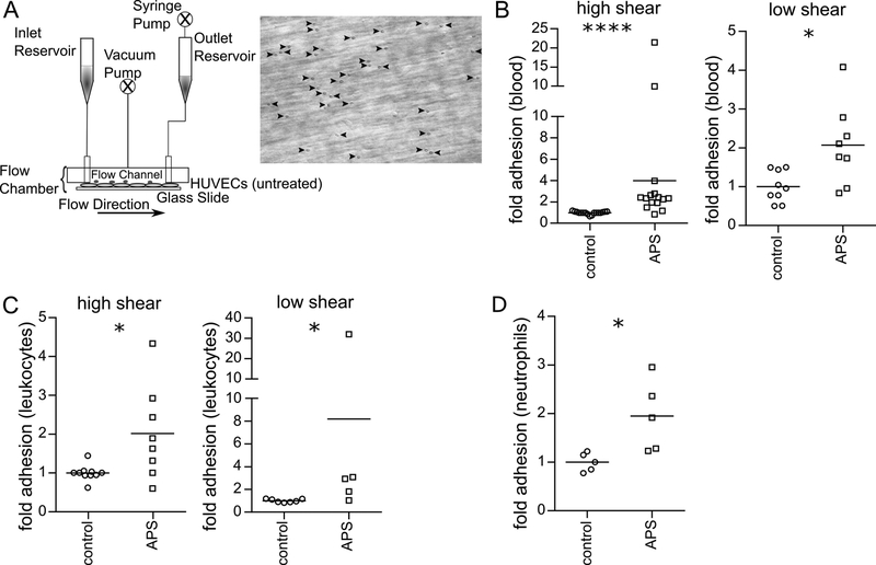
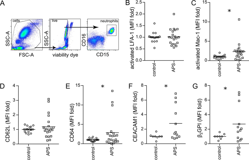
Methods: Patients with primary APS (n = 43), patients with a history of venous thrombosis but negative test results for antiphospholipid antibodies (n = 11), and healthy controls (n = 38) were studied. Cells were introduced into a flow chamber and perfused across resting human umbilical vein endothelial cells (HUVECs). Surface adhesion molecules were quantified by flow cytometry. Neutrophil extracellular trap release (NETosis) was assessed in neutrophil-HUVEC cocultures.

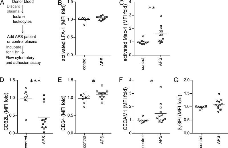
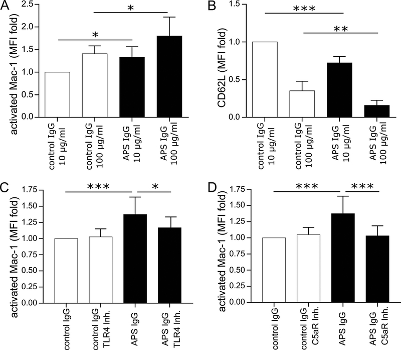
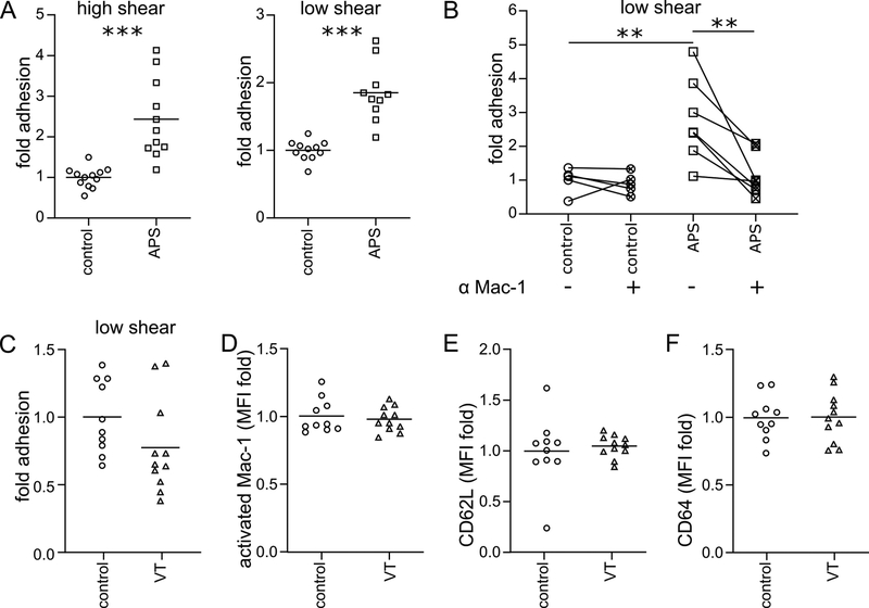
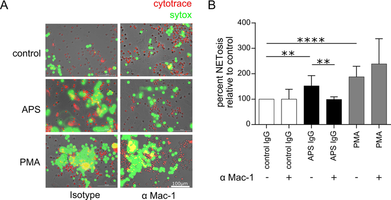
Results: Upon perfusion of anticoagulated blood through the flow chamber, APS neutrophils demonstrated increased adhesion as compared to control neutrophils under conditions representative of either venous (n = 8; P < 0.05) or arterial (n = 15; P < 0.0001) flow. At the same time, APS neutrophils were characterized by up-regulation of CD64, CEACAM1, β2 -glycoprotein I, and activated Mac-1 on their surface (n = 12-18; P < 0.05 for all markers). Exposing control neutrophils to APS plasma or APS IgG resulted in increased neutrophil adhesion (n = 10-11; P < 0.0001) and surface marker up-regulation as compared to controls. A monoclonal antibody specific for activated Mac-1 reduced the adhesion of APS neutrophils in the flow-chamber assay (P < 0.01). The same monoclonal antibody reduced NETosis in neutrophil-HUVEC cocultures (P < 0.01).

Conclusion: APS neutrophils demonstrate increased adhesive potential, which is dependent upon the activated form of Mac-1. In patients, this could lower the threshold for neutrophil-endothelium interactions, NETosis, and possibly thrombotic events.


Conflict of interest statement

AUTHORSHIP AND CONFLICT OF INTEREST DISCLOSURES

The authors have no competing interests or conflicts to disclose. GS, WJK, KG, SY, APV and ALB conducted experiments and analyzed data. GS, WJK, PLB, OE-A, and JSK designed the study. All authors participated in writing the manuscript, and gave approval before submission.

Conflict of interest: None of the authors has any financial conflict of interest to disclose.

Figures

Figure 1:
Figure 1:. APS neutrophils demonstrate increased adhesion.
Adhesion was measured under either pulsatile, high-shear (1000 s−1) conditions or laminar, low-shear (200 s−1) conditions. A, Schematic of the parallel-plate flow chamber, and a representative image from the adhesion assay. B, Anticoagulated whole blood from healthy controls or patients with APS were perfused through the flow chamber. At the end of the run, adherent cells were quantified; ****p<0.0001 (Mann Whitney test) and *p<0.05 (t test). C, Control or APS leukocytes were isolated, resuspended in flow buffer (plasma discarded), and perfused through the flow chamber; *p<0.05 (Mann Whitney test). D, Control or APS neutrophils were isolated, resuspended in flow buffer, and perfused through the flow chamber; *p<0.05 (t test).
Figure 2:
Figure 2:. Increased expression of activated Mac-1 and other adhesion molecules on APS neutrophils.
Flow cytometry was performed after treating anticoagulated whole blood with fluorescently-labeled antibodies. Mean fluorescence intensity (MFI) was normalized to controls run in the same batch. A, Schematic of gating strategy for identification of neutrophils in whole blood. B, Activated LFA-1 (not significant by t test). C, Activated Mac-1 (*p<0.05 by t test). D, CD62L (not significant by t test). E, CD64 (*p<0.05 by t test). F, CEACAM1 (*p<0.05 by t test). G, Beta-2 glycoprotein I (*p<0.05 by t test).
Figure 3:
Figure 3:. Increased expression of activated Mac-1 and other adhesion molecules when control neutrophils are conditioned with APS plasma.
Control leukocytes were conditioned with heterologous control plasma or APS plasma (A), and then incubated with fluorescently-labeled antibodies. Mean fluorescence intensity (MFI) was normalized to controls run in the same batch. B, Activated LFA-1 (not significant by t test). C, Activated Mac-1 (**p<0.01 by t test). D, CD62L (***p<0.001 by t test). E, CD64 (*p<0.05 by t test). F, CEACAM1 (*p<0.05 by Mann Whitney test). G, Beta-2 glycoprotein I (not significant by t test).
Figure 4:
Figure 4:. Exposure to purified APS IgG increases the expression of activated Mac-1 on control neutrophils in TLR4- and complement-dependent fashion.
Control leukocytes were treated with control or APS IgG as indicated. Activated Mac-1 and CD62L were quantified by flow cytometry. A, Activated Mac-1 (*p<0.05 by one-way ANOVA with correction for multiple comparisons by Holm-Sidak method; n=4 independent experiments). B, Shedding of CD62L (**p<0.01 and ****p<0.0001 by one-way ANOVA with correction for multiple comparisons by Holm-Sidak method; n=4 independent experiments). C, Control leukocytes were treated with control or APS IgG (100 μg/mL) in the presence or absence of TLR4 inhibitor. Activated Mac-1 was quantified by flow cytometry (*p<0.05 and ***p<0.001 by one-way ANOVA with correction for multiple comparisons by Holm-Sidak method; n=8 independent experiments). D, Control leukocytes were treated with control or APS IgG (100 μg/mL) in the presence or absence of C5a receptor (C5aR) inhibitor. Activated Mac-1 was quantified by flow cytometry (***p<0.001 by one-way ANOVA with correction for multiple comparisons by Holm-Sidak method; n=7 independent experiments).
Figure 5:
Figure 5:. Increased adhesion of APS leukocytes is mediated by activated Mac-1.
A, Control leukocytes were incubated with heterologous control or APS plasma, resuspended in flow buffer, and perfused through the flow chamber. Adherent cells were quantified (***p<0.001 by t test). B, Conditions were similar to panel A, except with the addition of a blocking antibody for activated Mac-1 to some samples (**p<0.01 by one-way ANOVA with correction for multiple comparisons by Holm-Sidak method). C, Similar to panel A, control leukocytes were incubated with heterologous control plasma or plasma from patients with history of unprovoked venous thrombosis (VT) but negative antiphospholipid testing. The leukocytes were then resuspended in flow buffer and perfused through the flow chamber. Adherent cells were quantified (not significant by t test). D-F, Similar to Figure 3, control leukocytes were conditioned with heterologous control plasma or VT plasma, and then incubated with fluorescently-labeled antibodies. Mean fluorescence intensity (MFI) was normalized to controls run in the same batch. D, Activated Mac-1 (not significant by t test). E, CD62L (not significant by t test). F, CD64 (not significant by t test).
Figure 6:
Figure 6:. Activated Mac-1 regulates APS IgG-mediated NETosis.
Control neutrophils were plated over a monolayer of HUVECs in the presence of either activated-Mac-1 blocking antibody or isotype control. Cultures were then stimulated with control IgG (100 μg/mL), APS IgG (100 μg/mL), or phorbol myristate acetate (PMA, positive control). After 3 hours, SYTOX Green was added to the culture, and fluorescence intensity was quantified. A, In these representative images, live cells are labeled by CytoTrace™ Red and extracellular DNA (NETs) by SYTOX Green; scale bar=100 μm. B, Quantification of NETosis by fluorescence intensity of SYTOX Green (**p<0.01 and ****p<0.0001 by one-way ANOVA with correction for multiple comparisons by Holm-Sidak method; n=4 independent experiments).

References

    1. Miyakis S, Lockshin MD, Atsumi T, Branch DW, Brey RL, Cervera R, et al. International consensus statement on an update of the classification criteria for definite antiphospholipid syndrome (APS). J Thromb Haemost. 2006;4(2):295–306. - PubMed
    1. Garcia D, Erkan D. Diagnosis and Management of the Antiphospholipid Syndrome. N Engl J Med. 2018;378(21):2010–21. - PubMed
    1. Abreu MM, Danowski A, Wahl DG, Amigo MC, Tektonidou M, Pacheco MS, et al. The relevance of “non-criteria” clinical manifestations of antiphospholipid syndrome: 14th International Congress on Antiphospholipid Antibodies Technical Task Force Report on Antiphospholipid Syndrome Clinical Features. Autoimmun Rev. 2015;14(5):401–14. - PubMed
    1. Rao AN, Kazzaz NM, Knight JS. Do neutrophil extracellular traps contribute to the heightened risk of thrombosis in inflammatory diseases? World J Cardiol. 2015;7(12):829–42. - PMC - PubMed
    1. Yalavarthi S, Gould TJ, Rao AN, Mazza LF, Morris AE, Nunez-Alvarez C, et al. Release of neutrophil extracellular traps by neutrophils stimulated with antiphospholipid antibodies: a newly identified mechanism of thrombosis in the antiphospholipid syndrome. Arthritis Rheumatol. 2015;67(11):2990–3003. - PMC - PubMed

Publication types

Substances