. 2019 Jun 11:12:4555-4566.
doi: 10.2147/OTT.S210623. eCollection 2019.

Pseudogene DUXAP10 acts as a diagnostic and prognostic marker and promotes cell proliferation by activating PI3K/AKT pathway in hepatocellular carcinoma

Affiliations

Pseudogene DUXAP10 acts as a diagnostic and prognostic marker and promotes cell proliferation by activating PI3K/AKT pathway in hepatocellular carcinoma

Chaosen Yue et al. Onco Targets Ther. .

Abstract

Background: Recently, the pseudogene DUXAP10 was shown to be overexpressed in various human cancers and emerged as a key cancer regulator. However, the roles of DUXAP10 in hepatocellular carcinoma (HCC) tumorigenesis and progression remain uncharacterized. Methods: Comprehensive analyses were performed to investigate DUXAP10 expression patterns, potential biologic functions, and clinical significance in HCC based on the data downloaded from the Cancer Genome Atlas (TCGA) and Gene Expression Omnibus (GEO) databases. DUXAP10 expression levels in HCC tissue sections and cells were verified using quantitative real-time PCR analysis. DUXAP10-siRNA was used to silence DUXAP10 in the Hep3B cell line to determine the roles of DUXAP10 in HCC cell proliferation. Results: DUXAP10 was significantly overexpressed in HCC, and DUXAP10 upregulation was closely associated with poor prognoses in HCC patients. DUXAP10 knockdown decreased cell proliferation and arrested HCC cells in the G1 phase of the cell cycle. Western blot analysis showed that DUXAP10 knockdown decreased p-AKT expression in HCC cells. Conclusion: Our study demonstrates that pseudogene DUXAP10 promotes HCC cell proliferation by activating PI3K/AKT pathway and could act as a potential diagnostic and prognostic biomarker for HCC patients.

Keywords: DUXAP10; HCC; biomarker.


Conflict of interest statement

The authors report no conflicts of interest in this work.

Figures
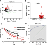
Figure 1
Figure 1
DUXAP10 expression patterns in HCC based on TCGA database. (A) Volcano plot of differentially expressed pseudogenes screened based on TCGA database (|log2FC|>2 and P-value<0.01). (B) DUXAP10 expression levels in HCC tissues compared with adjacent liver tissues of TCGA. (C) Overall survival plots of DUXAP10 in TCGA cohort. (D) Significance of DUXAP10 in HCC with the ROC curve analysis from TCGA. Abbreviations: HCC, hepatocellular carcinoma; TCGA, the Cancer Genome Atlas; FC, fold change.
Figure 2
Figure 2
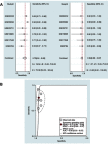
Forest plot of studies evaluating the DUXAP10 expression level between hepatocellular carcinoma tissues and adjacent liver tissues. Meta-analysis was used to calculate the pooled SMD with 95% CI. Abbreviations: SMD, standard mean difference; CI, confidence interval.
Figure 3
Figure 3
Diagnostic value of DUXAP10 in HCC based on multiple GEO datasets. (A) Pooled sensitivity and specificity of DUXAP10 expression for diagnosis of HCC. (B) Pooled AUC was calculated by SROC curve analysis. Abbreviations: HCC, hepatocellular carcinoma; GEO, Gene Expression Omnibus; AUC, area under the curve; SROC, Summary receiver operating characteristic curve.
Figure 4
Figure 4
GO (A) and KEGG pathway (B) enrichment analysis of the DUXAP10-related protein-coding genes. Abbreviations: GO, Gene Ontology; KEGG, Kyoto Encyclopedia of Genes and Genomes.
Figure 5
Figure 5
Verification of DUXAP10 upregulation in HCC via qRT-PCR analysis. (A) DUXAP10 expression levels of 46 HCC tissues samples and matched adjacent liver tissues samples were measured by qRT-PCR. (B) Overall survival plots of DUXAP10 based on the 46 HCC patients’ clinical data. (C) DUXAP10 expression levels in HCC cell lines and normal liver cells were measured by qRT-PCR. Abbreviations: HCC, hepatocellular carcinoma; qRT-PCR, quantitative real-time polymerase chain reaction.
Figure 6
Figure 6
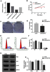
Silencing DUXAP10 inhibited Hep3B cells proliferation through PI3K/AKT pathway (A) si-DUXAP10-2# had the highest interference efficiency according to qRT-PCR analysis. (B) Cell viability was detected by the CCK-8 assay. (C and D) DUXAP10 knockdown inhibited the colony formation ability of HCC cells. (E and F) DUXAP10 knockdown arrested HCC cells in the G1 phase of the cell cycle. (G and H) DUXAP10 knockdown decreased p-AKT protein expression in HCC cells. All experiments in this study were performed in triplicate and the results were represented as the mean±SD. *P<0.05. Abbreviations: qRT-PCR, quantitative real-time polymerase chain reaction; CCK-8, Cell Counting Kit-8.

References

    1. Siegel RL, Miller KD, Jemal A. Cancer statistics, 2018. CA Cancer J Clin. 2018;68(1):7–30. doi:10.3322/caac.21442 - DOI - PubMed
    1. Chen W, Zheng R, Baade PD, et al. Cancer statistics in China, 2015. CA Cancer J Clin. 2016;66(2):115–132. doi:10.3322/caac.21338 - DOI - PubMed
    1. Tang A, Hallouch O, Chernyak V, Kamaya A, Sirlin CB. Epidemiology of hepatocellular carcinoma: target population for surveillance and diagnosis. Abdom Radiol (NY). 2018;43(1):13–25. doi:10.1007/s00261-017-1209-1 - DOI - PubMed
    1. Kulik L, El-Serag HB. Epidemiology and Management of Hepatocellular Carcinoma. Gastroenterology. 2019;156(2):477–491. doi:10.1053/j.gastro.2018.08.065 - DOI - PMC - PubMed
    1. Proudfoot N. Pseudogenes. Nature. 1980;286:840–841. - PubMed