

Vascular index measured by smart 3-D superb microvascular imaging can help to differentiate malignant and benign breast lesion
- PMID: 31354354
- PMCID: PMC6580120
- DOI: 10.2147/CMAR.S203376
Vascular index measured by smart 3-D superb microvascular imaging can help to differentiate malignant and benign breast lesion
Abstract
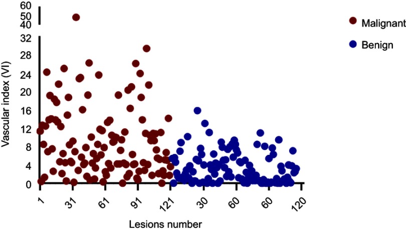
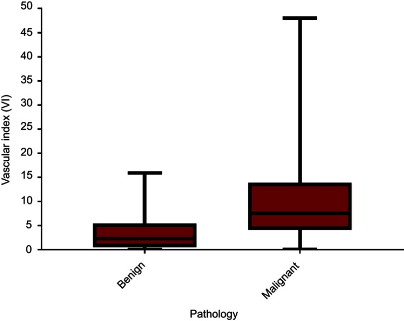
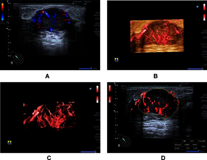
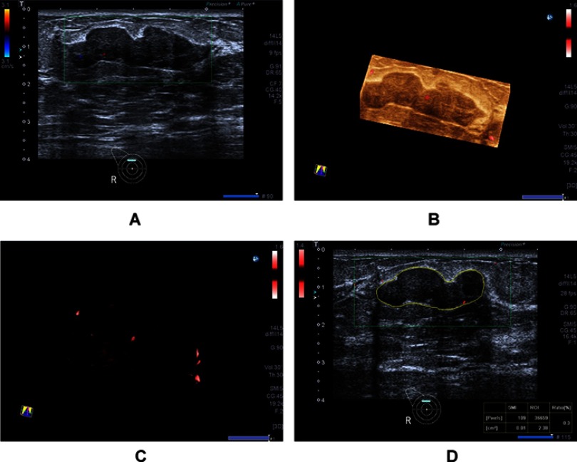
Purpose: The purpose of our study was to prospectively evaluate the diagnostic performance of the vascular index (VI, defined as the ratio of Doppler signal pixels to pixels in the total lesion) measured via Smart 3-D superb microvascular imaging (SMI) for breast lesions. Patients and methods: Two hundred and thirty-two consecutive patients with 236 breast lesions referred for biopsy at Peking Union Medical College Hospital were enrolled in the study from December 2016 to November 2017. Sensitivity, specificity, positive predictive value (PPV), negative predictive value (NPV) and accuracy of VI were calculated with histopathologic results as the reference standard. Results: Of the 236 breast lesions, 121 were malignant and 115 were benign. The mean VI was significantly higher in malignant lesions (9.7±8.2) than that in benign ones (3.4±3.3) (P<0.0001). Sensitivity, specificity, PPV, NPV and accuracy of VI (4.0 as the threshold) were respectively: 76.0%, 66.1%, 70.2%, 72.4% and 71.2% (P<0.05). Conclusion: Smart three-dimensional (3-D) SMI is a noninvasive tool using two-dimensional (2-D) scanning to generate 3-D vascular architecture with a high-resolution image of micro-vessels. This can be used as a qualitative guide to identify the optimal 2-D SMI plane with the most abundant vasculature to guide VI quantitative measurements of breast lesions. Smart 3-D SMI may potentially serve as a noninvasive tool to accurately characterize benign versus malignant breast lesions.
Keywords: breast neoplasms; diagnostic imaging; superb microvascular imaging; ultrasonography.
Conflict of interest statement
The authors report no conflicts of interest in this work.
Figures
References
-
- Less JR, Skalak TC, Sevick EM, Jain RK. Microvascular architecture in a mammary carcinoma: branching patterns and vessel dimensions. Cancer Res. 1991;51(1):265–273. - PubMed
LinkOut - more resources
Full Text Sources
