Review
. 2019 Jul 11:10:705.
doi: 10.3389/fphys.2019.00705. eCollection 2019.

Modeling Mammalian Commitment to the Neural Lineage Using Embryos and Embryonic Stem Cells

Affiliations
Review

Modeling Mammalian Commitment to the Neural Lineage Using Embryos and Embryonic Stem Cells

Rachel A Shparberg et al. Front Physiol. .

Abstract

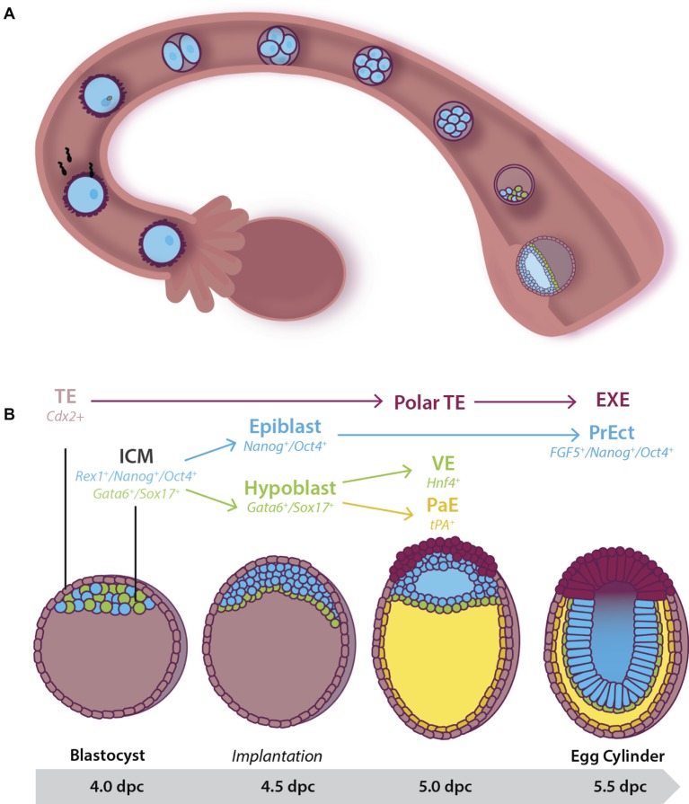
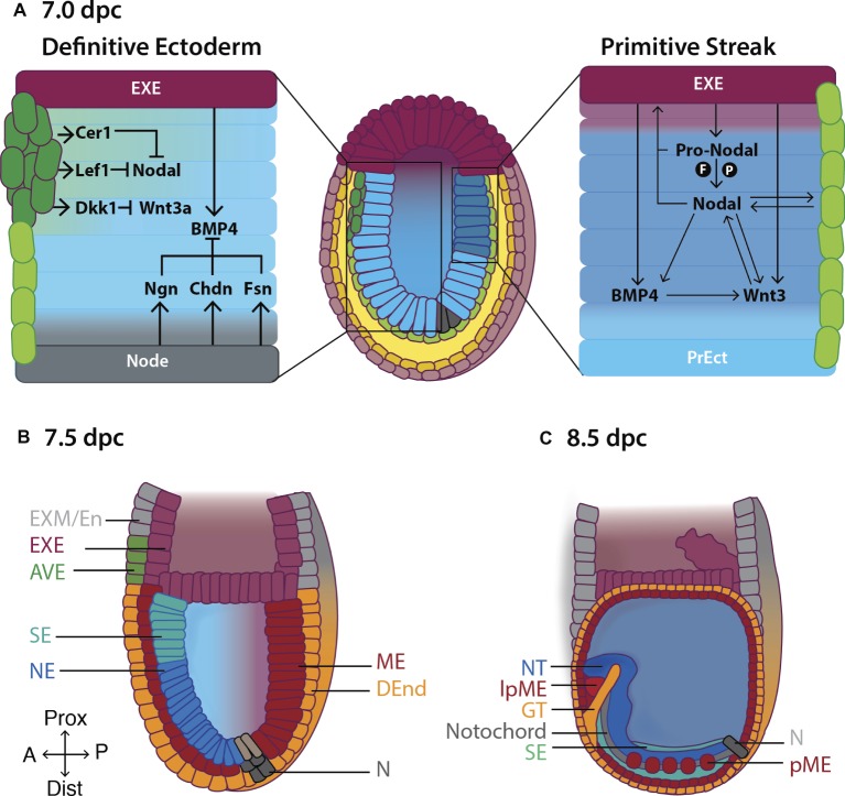
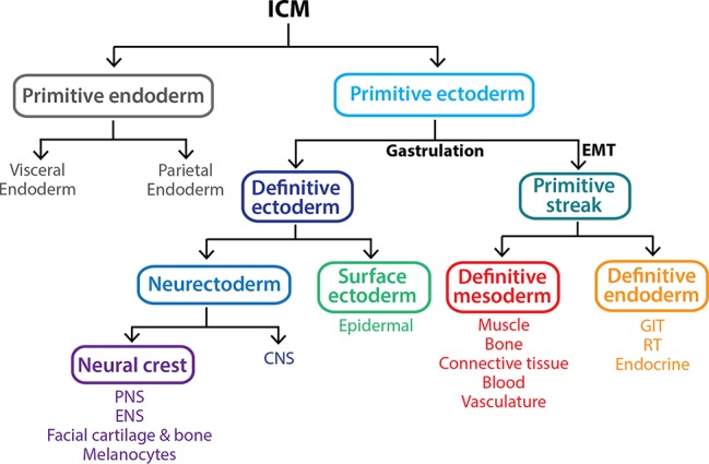
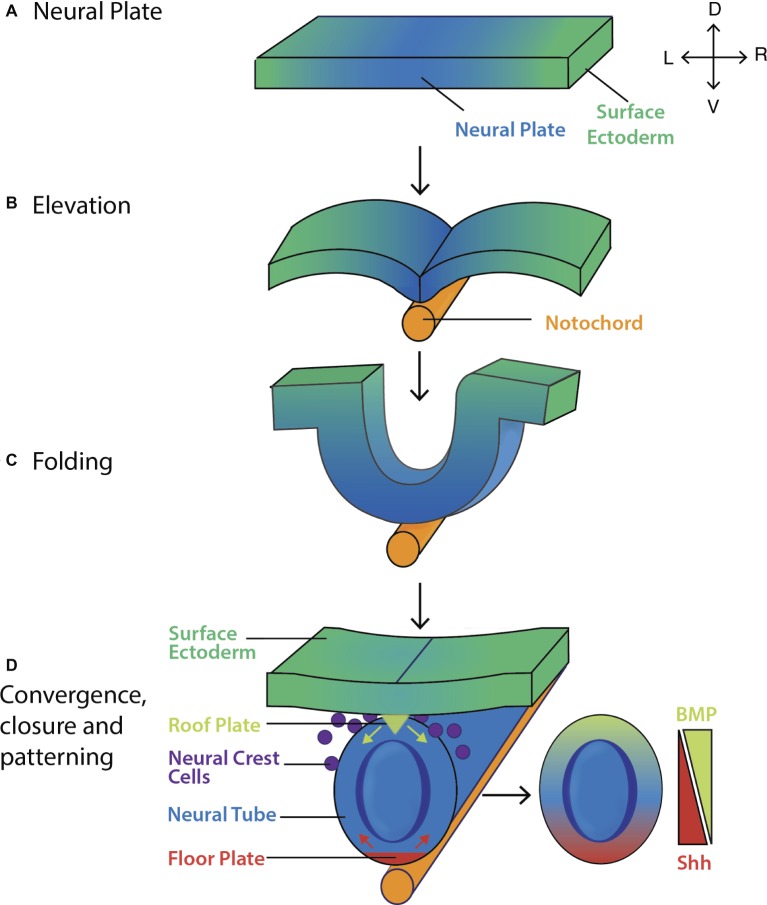
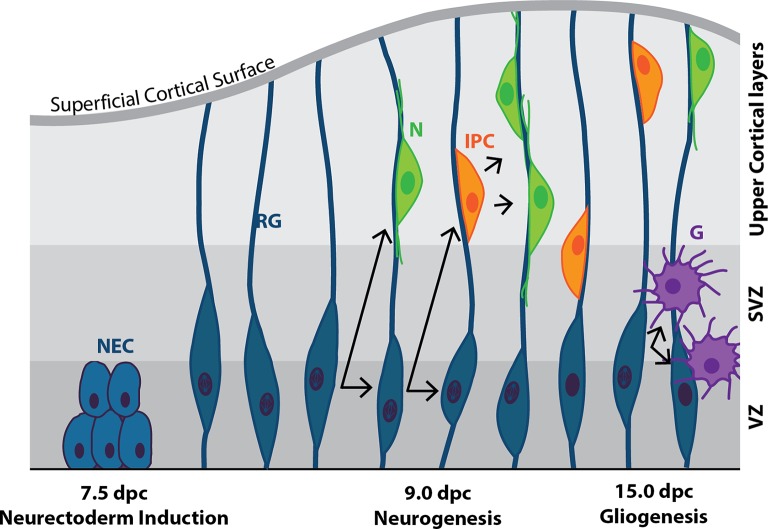
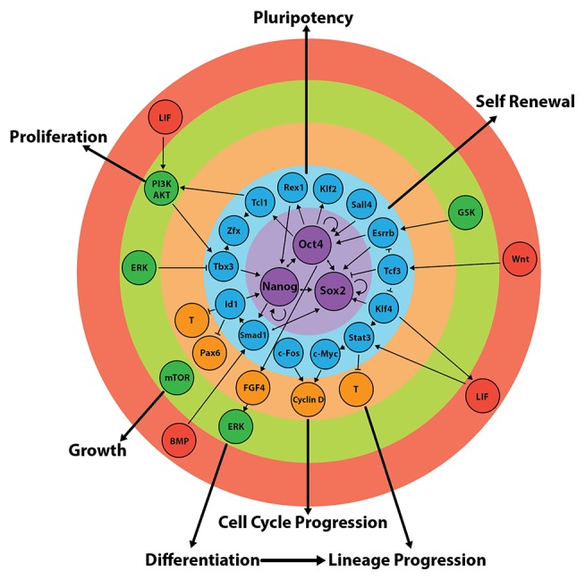
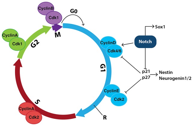
Early mammalian embryogenesis relies on a large range of cellular and molecular mechanisms to guide cell fate. In this highly complex interacting system, molecular circuitry tightly controls emergent properties, including cell differentiation, proliferation, morphology, migration, and communication. These molecular circuits include those responsible for the control of gene and protein expression, as well as metabolism and epigenetics. Due to the complexity of this circuitry and the relative inaccessibility of the mammalian embryo in utero, mammalian neural commitment remains one of the most challenging and poorly understood areas of developmental biology. In order to generate the nervous system, the embryo first produces two pluripotent populations, the inner cell mass and then the primitive ectoderm. The latter is the cellular substrate for gastrulation from which the three multipotent germ layers form. The germ layer definitive ectoderm, in turn, is the substrate for multipotent neurectoderm (neural plate and neural tube) formation, representing the first morphological signs of nervous system development. Subsequent patterning of the neural tube is then responsible for the formation of most of the central and peripheral nervous systems. While a large number of studies have assessed how a competent neurectoderm produces mature neural cells, less is known about the molecular signatures of definitive ectoderm and neurectoderm and the key molecular mechanisms driving their formation. Using pluripotent stem cells as a model, we will discuss the current understanding of how the pluripotent inner cell mass transitions to pluripotent primitive ectoderm and sequentially to the multipotent definitive ectoderm and neurectoderm. We will focus on the integration of cell signaling, gene activation, and epigenetic control that govern these developmental steps, and provide insight into the novel growth factor-like role that specific amino acids, such as L-proline, play in this process.

Keywords: L-proline; amino acids; definitive ectoderm; early primitive ectoderm-like cells; embryonic stem cell; neural; neurectoderm; primitive ectoderm.


Figures

Figure 1
Figure 1
Early pre- and post-implantation mouse embryonic development until the egg cylinder stage. (A) Following fertilization, the embryo undergoes a series of cleavage divisions as it travels down the fallopian tube. Between 3.5 and 4.5 dpc, the embryo, now known as a blastocyst, consists of two cell populations: An outer multipotent trophectoderm (TE) (expressing Cdx2), and a mosaic inner pluripotent inner cell mass (ICM) population. At 4.0 dpc, the blastocyst hatches from the zona pelucida and implants into the uterine wall. (B) Cells of the 4.0 dpc ICM expressing Gata6 and Sox17 move to line the blastocoelic cavity, lose pluripotency, and differentiate into the extraembryonic primitive endoderm (or hypoblast) by 4.5 dpc. Together the remaining cells of the ICM (the epiblast) and the hypoblast form the bilaminar disc by 5.0 dpc. At this stage, cells of the pluripotent epiblast that have not moved to be in contact with the extracellular matrix laid down between the hypoblast undergo apoptosis to help form the proamniotic cavity. Hypoblast cells that remain in close contact with the epiblast differentiate into visceral endoderm (VE) while those that migrate along the basement membrane of the TE form the tPA+ parietal endoderm (PaE), resulting in the formation of the yolk sac. At 5.5 dpc the embryo is known as the egg cylinder. The remaining surviving epiblast cells have differentiated into a second pluripotent population of pseudo-stratified cells known as the primitive ectoderm (PrEct). The TE differentiates into cells that constitute the placenta including the extraembryonic endoderm (EXE).
Figure 2
Figure 2
Formation of the primitive body plan following gastrulation in the mouse. (A) Right hand panel: Pro-Nodal secreted from the extraembryonic ectoderm (EXE) is converted to Nodal in the presence of the convertases Furin (F) and Pace4 (P). Nodal acts on the visceral endoderm (light green cells) to regulate the expression of pro-Nodal and production of Nodal. A feedback system is established between Nodal, BMP and Wnt3 causing primitive ectoderm (PrEct) cells on the posterior side to ingress through the primitive streak, which continues to elongate in a proximal-distal direction from 6.5 dpc. Cells that migrate through the primitive streak form the definitive mesoderm (ME) and endoderm (DEnd) germ layers. Left hand panel: On the anterior side of the embryo, the anterior visceral endoderm (AVE; dark green cells) secretes the Nodal antagonists Cer1 and Lef1, and the Wnt3 antagonist Dkk1, inhibiting Nodal and Wnt3 signaling and thus establishing the definitive ectoderm germ layer by 7.0 dpc. BMP4 is secreted from the EXE, while BMP4 antagonists including Noggin (Ngn), Chordin (Chd) and Follistatin (Fsn) are secreted from the Node (N), establishing a gradient of BMP4 across the definitive ectoderm, such that by (B) 7.5 dpc, BMP4-mediated SMAD signaling in the proximal definitive ectoderm produces surface ectoderm (SE) while the distal definitive ectoderm differentiates to neurectoderm (NE) in the absence of SMAD signaling. (C) Following the completion of gastrulation at ~7.5 dpc, the ME differentiates to give rise to the paraxial mesoderm (pME) and lateral plate mesoderm (lpME), the DEnd produces the gut tube (GT) and the NE gives rise to the neural tube (NT). Additional key: EXM/En, extraembryonic mesoderm/endoderm; Prox, proximal; Dist, distal; A, anterior; P, posterior.
Figure 3
Figure 3
Gastrulation gives rise to the three primary germ layers of the embryo proper. The inner cell mass (ICM) is a pluripotent population of cells that arises between 3.5 and 4.5 dpc within the blastocyst. By 5.5 dpc, the ICM differentiates into the multipotent extraembryonic primitive endoderm lineage and a second pluripotent population, the primitive ectoderm. At 6.5 dpc, the primitive ectoderm undergoes gastrulation in response to various signals including Nodal, resulting in a subset of cells undergoing an epithelial-to-mesenchymal transition (EMT), allowing them to ingress through the primitive streak and form the definitive mesoderm and definitive endoderm germ layers. The remaining primitive ectoderm cells (which see little or no Nodal) do not move through the streak and give rise to the definitive ectoderm germ layer, which further differentiates into the surface ectoderm and neurectoderm in response to the presence and absence of BMP4 signaling, respectively. Key: CNS, central nervous system; ENS, enteric nervous system; GIT, gastrointestinal tract (epithelial lining); PNS, peripheral nervous system; RT, respiratory tract (epithelial lining).
Figure 4
Figure 4
Formation and patterning of the mouse neural tube. (A) The pseudostratified columnar epithelium of neural plate forms by 7.5 dpc. The lateral edges of the neural plate then (B) elevate and (C) fold by ~8.0 dpc before (D) converging at the midline and closing by ~8.5 dpc. Shh (red arrows) and BMP inhibitors secreted from the floor plate, and BMP4/7 (green arrows) secreted from the roof plate act to pattern the neural tube along its ventro-dorsal axis, giving rise to the layers of the spinal cord (Gilbert, 2006). Key: V, ventral; D, dorsal; L, left; R, right.
Figure 5
Figure 5
Cortical neurogenesis in the mouse. Following neural plate formation at 7.5 dpc, neuroepithelial cells (NEC) differentiate into mitotically active neural progenitor cells known as radial glia (RG) by ~9.0 dpc. RG undergo either symmetrical division to produce two RG daughter cells, or asymmetric division to produce one RG daughter cell and a terminally-differentiated neuron (N), or an intermediate progenitor cell (IPC), or a mature glial cell (G). IPCs are capable of undergoing symmetrical division to form neurons. N and IPCs migrate along the axons of the RG cells from the ventricular zone (VZ), through the subventricular zone (SVZ) and into the upper cortical layers of the developing brain.
Figure 6
Figure 6
Regulation of the core and extended pluripotency networks in mouse embryonic stem cells. Maintenance of pluripotency and self-renewal is governed by external stimuli (red), which act on various signalling pathways (green) that regulate the expression of the extended (blue) and core (purple) pluripotency networks. In turn, the expression of transcription factors of the core circuitry regulates their own expression, as well as the expression of other factors involved in differentiation and/or self-renewal (orange). Figure adapted from data published in: (Jirmanova et al., 2002; Mitsui et al., 2003; Ying et al., 2003a; Paling et al., 2004; Hamazaki et al., 2006; Binétruy et al., 2007; Kunath et al., 2007; Storm et al., 2007, 2009; Chen et al., 2008; Medvedev et al., 2008; Niwa et al., 2009; Hall et al., 2009b; Hirai et al., 2011; Wray et al., 2011; Kim et al., 2012; Marks et al., 2012; Nichols and Smith, 2012; Romero-Lanman et al., 2012; Do et al., 2013; Lee, 2013; Hamilton and Brickman, 2014; Posfai et al., 2014; Tosolini and Jouneau, 2015).
Figure 7
Figure 7
Cell signalling events that mediate the switch between self-renewal and differentiation in mouse embryonic stem cells. (A) LIF binds to the LIF receptor (LIFR) resulting in heterodimerisation with glycoprotein-130 (gp130). Downstream JAK proteins become phosphorylated, and in this active state phosphorylate tyrosine residues on the receptor complex. STAT3 can then dock to the receptor, is then phosphorylated at Y705, and homodimerises before translocating to the nucleus where it induces the transcription of self-renewal genes. LIF also activates the PI3K pathway, in which PIP2 is converted to PIP3 resulting in the downstream phosphorylation of AKT at S473 and T308. This enhances self-renewal by upregulating the expression of Nanog, and by promoting cell cycle progression. (B) BMP4 binds to its cognate BMP receptor (BMPR) resulting in the phosphorylation of SMAD1/5/8. Once phosphorylated, SMAD1/5/8 forms heterodimers with SMAD4, which translocate to the nucleus resulting in transcription of inhibitor-of-differentiation (Id) genes. SMAD signaling also results in the upregulation of the phosphatase DUSP9 which acts as a negative regulator of ERK, thereby inhibiting differentiation. (C) LIF also activates the MAPK/ERK pathway to promote differentiation in the face of maintaining self-renewal. The balance can be tipped toward differentiation by the presence of Fibroblast Growth Factor (FGF4), which upregulates the activity of the MAPK/ERK pathways, as does (D) L-proline. L-proline enters the cell via the Sodium-coupled Neutral Amino Acid Transporter (SNAT)-2 where it activates mTOR to induce the differentiation of mESCs to early primitive ectoderm-like (EPL) cells. Figure adapted from data published in: (Ying et al., 2003b; Paling et al., 2004; Binétruy et al., 2007; Washington et al., 2010; Hirai et al., 2011; Romero-Lanman et al., 2012; Hamilton and Brickman, 2014).
Figure 8
Figure 8
Notch signaling in neural stem cells promotes cell-cycle progression and inhibits neurogenesis. During the mammalian cell cycle, the length of time it takes cells to move through interphase (G1, S and G2) and, in particular, how long they spend in G1, helps determine their fate. Transition between stages of the cell cycle is driven by a series of cyclin-dependent kinases (Cdks) binding to specific cyclins to promote cell-cycle progression. Notch signaling increases the expression of both CyclinD and Cdk4/6, indirectly allowing cells to progress past the restriction checkpoint (R) and into S phase, promoting proliferation. Notch also inhibits the cell-cycle inhibitors p21 and p27, which further promotes proliferation and prevents the expression of genes involved in neurogenesis such as Nestin and Neurogenin1/2.
Figure 9
Figure 9
Model of L-proline-mediated differentiation of mESCs to EPL cells. L-Pro enters the cell via the SNAT2 transporter (Tan et al., 2011) where it (A) acts on various signaling pathways including the amino acid-sensing signaling mTOR pathway, ERK and P38 pathways to induce differentiation (Lonic, 2007; Washington et al., 2010; Tan et al., 2016). (B) L-Pro is metabolized to pyrroline-5-carboxylate (P5C) by proline oxidase (POX) via the proline cycle in the mitochondria. P5C can then be converted to glutamate (Glu) by P5C dehydrogenase (P5CD). Glu is further converted to alpha-ketoglutarate, which enters the TCA cycle with subsequent production of ATP via oxidative phosphorylation (OxPhos). Glu can also be converted to L-Pro via the sequential actions of P5C synthase (P5CS) and P5C reductase (P5CR). This conversion produces NADP+, which is required to stimulate the pentose phosphate pathway (PPP) in the cytoplasm of the cell (Pandhare et al., 2009; Phang et al., 2010). The PPP is required to generate the ribose components needed for DNA and RNA synthesis and is coupled with aerobic glycolysis to generate ATP in highly proliferative cells such as ESCs and cancer cells (Liu et al., 2015). Stimulation of the PPP and glycolysis in the presence of L-Pro (Liu et al., 2015) may stimulate the differentiation of mESCs to EPL cells and support the increased proliferation rates observed in EPL cells. (C) When mESCs are starved of L-Pro, the amino acid response (AAR) pathway is activated to control L-Pro production and uptake into the cell. Free prolyl-tRNA (i.e., not loaded with L-Pro) binds to General Control Non-Depressible-2 (Gcn2) kinase which phosphorylates Eukaryotic Initiation Factor-2 (eIF2), resulting in Activating Transcription Factor-4 (Atf4) regulating the expression of genes involved in L-Pro transport and metabolism. The net result of this autoregulatory loop is self-limiting concentrations of L-Pro resulting in mESC self-renewal (D’Aniello et al., 2015). When L-Pro is added exogenously, this pathway is overwhelmed triggering differentiation.

Similar articles

Cited by

References

    1. Aguilar J., Reyley M. (2005). The uterine tubal fluid: secretion, composition and biological effects. Anim. Reprod. 2, 91–105. Available at: http://www.cbra.org.br/pages/publicacoes/animalreproduction/issues/downl... [Accessed September 10, 2015]
    1. Alarcon V. B. (2010). Cell polarity regulator PARD6B is essential for trophectoderm formation in the preimplantation mouse embryo. Biol. Reprod. 83, 347–358. 10.1095/biolreprod.110.084400 - DOI - PMC - PubMed
    1. Anderson C., Stern C. D. (2016). Organizers in development. Curr. Top. Dev. Biol. 117, 435–454. 10.1016/bs.ctdb.2015.11.023 - DOI - PubMed
    1. Ang S. L., Rossant J. (1994). HNF-3 beta is essential for node and notochord formation in mouse development. Cell 78, 561–574. 10.1016/0360-3016(94)90351-4, PMID: - DOI - PubMed
    1. Ang Y.-S., Tsai S.-Y., Lee D.-F., Monk J., Su J., Ratnakumar K., et al. (2011). Wdr5 mediates self-renewal and reprogramming via the embryonic stem cell core transcriptional network. Cell 145, 183–197. 10.1016/j.cell.2011.03.003, PMID: - DOI - PMC - PubMed

LinkOut - more resources