Review
. 2019 Jul 10:10:1490.
doi: 10.3389/fmicb.2019.01490. eCollection 2019.

RNA Synthesis and Capping by Non-segmented Negative Strand RNA Viral Polymerases: Lessons From a Prototypic Virus

Affiliations
Review

RNA Synthesis and Capping by Non-segmented Negative Strand RNA Viral Polymerases: Lessons From a Prototypic Virus

Tomoaki Ogino et al. Front Microbiol. .

Abstract

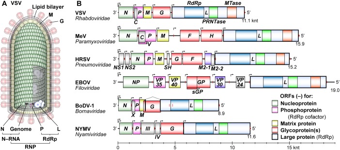
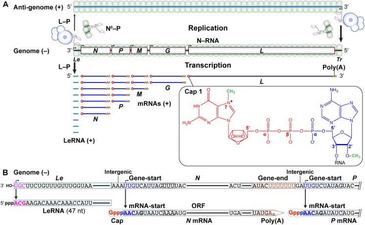
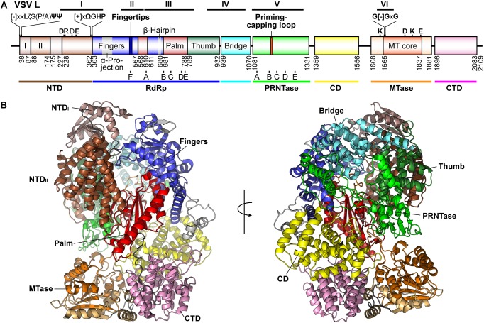
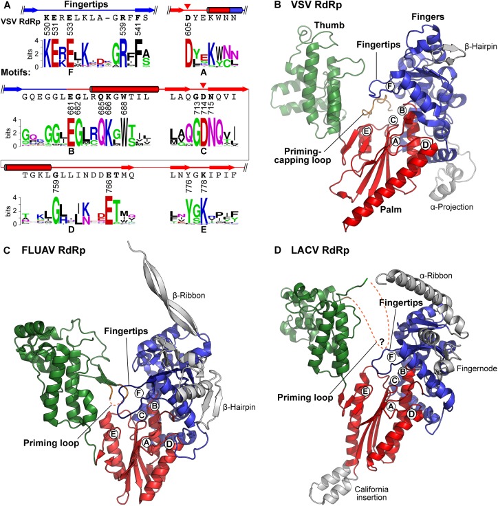
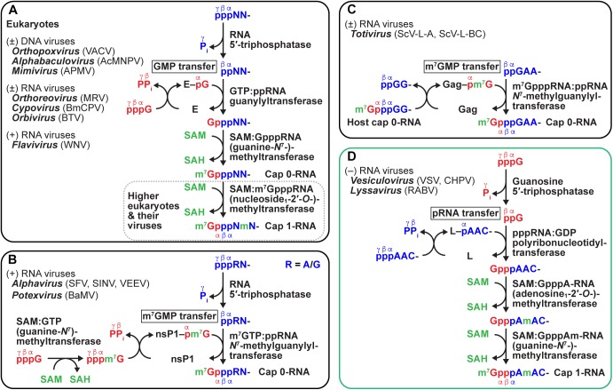
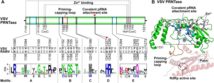
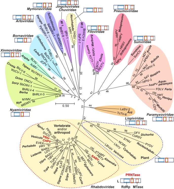
Non-segmented negative strand (NNS) RNA viruses belonging to the order Mononegavirales are highly diversified eukaryotic viruses including significant human pathogens, such as rabies, measles, Nipah, and Ebola. Elucidation of their unique strategies to replicate in eukaryotic cells is crucial to aid in developing anti-NNS RNA viral agents. Over the past 40 years, vesicular stomatitis virus (VSV), closely related to rabies virus, has served as a paradigm to study the fundamental molecular mechanisms of transcription and replication of NNS RNA viruses. These studies provided insights into how NNS RNA viruses synthesize 5'-capped mRNAs using their RNA-dependent RNA polymerase L proteins equipped with an unconventional mRNA capping enzyme, namely GDP polyribonucleotidyltransferase (PRNTase), domain. PRNTase or PRNTase-like domains are evolutionally conserved among L proteins of all known NNS RNA viruses and their related viruses belonging to Jingchuvirales, a newly established order, in the class Monjiviricetes, suggesting that they may have evolved from a common ancestor that acquired the unique capping system to replicate in a primitive eukaryotic host. This article reviews what has been learned from biochemical and structural studies on the VSV RNA biosynthesis machinery, and then focuses on recent advances in our understanding of regulatory and catalytic roles of the PRNTase domain in RNA synthesis and capping.

Keywords: GDP polyribonucleotidyltransferase; RNA-dependent RNA polymerase; mRNA capping; non-segmented negative strand RNA viruses; rabies virus; replication; transcription; vesicular stomatitis virus.


Figures

FIGURE 1
FIGURE 1
Schematic diagrams of a VSV virion and NNS RNA viral genomes. (A) A VSV particle is composed of a cellular lipid bilayer, viral RNA genome, and five viral proteins: nucleo- (N), phospho- (P), matrix (M), glyco- (G), and large (L) proteins. The virus particle contains a ribonucleoprotein (RNP) composed of N–RNA and RNA-dependent RNA polymerase (RdRp) complexes. (B) The gene organization of negative-strand genomes of typical NNS RNA viruses [measles virus (MeV), human respiratory syncytial virus (HRSV), Ebola virus (EBOV), Borna disease virus 1 (BoDV-1), and Nyamanini virus (NYMV)] belonging to different families is depicted in the 3′ to 5′ order. Transcription initiation and termination sites are shown by bent arrows and red vertical lines, respectively. The positions of negative-strand open reading frames are shown by colored boxes. The L genes encode an L protein with RdRp, GDP polyribonucleotidyltransferase (PRNTase), and methyltransferase (MTase, except for nuclear-replicating viruses) domains. A scale bar is shown at the bottom (knt, kilo nucleotides).
FIGURE 2
FIGURE 2
Transcription and replication of the VSV genome. (A) The negative-strand VSV genome in the N–RNA complex serves as a template for transcription (lower) and replication (upper). Le and Tr denote the terminal leader and trailer regions, respectively, in the genome. According to the single-entry, stop-start transcription model, the L–P RdRp complex enters from the 3′-end of the genome and sequentially synthesizes the leader RNA (LeRNA) and five monocistronic mRNAs with a 5′-cap 1 structure and 3′-poly(A) tail (lower). A GDP moiety (red) of GTP, an AMP moiety (blue) of ATP, and two methyl groups (green) are incorporated into the cap 1 structure. The L–P and N0–P (N0: RNA-free N) complexes are required for encapsidation-coupled genome replication (upper). (B) LeRNA is synthesized from the 3′-terminal of the Le promoter in the genome. The conserved gene-start and gene-end sequences serve as internal transcription initiation and termination/polyadenylation signals, respectively. The conserved 5′-terminal mRNA-start sequence acts as a signal for mRNA capping.
FIGURE 3
FIGURE 3
Structure of the VSV N protein. (A) The domain organization of the VSV N protein is schematically represented. Basic residues contributing to RNA binding are noted above the schematic, and residues involved in P-binding are noted with a blue bar. (B) A cartoon representation of the monomeric N protein (PDB id: 2GIC) is shown with bound nine-mer of RNA encapsidated and regional landmarks noted. (C) Assembled trimer of N proteins (each represented in a different color) with bound 27-mer of RNA is shown. All illustrations were prepared with PyMOL (DeLano, 2002).
FIGURE 4
FIGURE 4
Structure of the VSV P protein. (A) The domain organization of the VSV P protein is schematically represented. Domains are labeled according to known binding partners and function. Six phosphorylation sites are noted above the schematic. (B) VSV P exists as a dimer and is represented in cartoon form with regional aspects noted in (A) labeled. P binds the unassembled N0 (top) via a single helix and adjacent amino acids in the N-terminal intrinsically disordered region (PDB id: 3PMK). The dimerization domain (PDB id: 2FQM) is shown central to the figure. The C-terminal domain of P (PDB id: 3HHZ) binds a bipartite binding site involving the C-loops and an α-helix in the C-lobe of N. A tetramer of N proteins (each represented in a different color) is shown in surface representation. The view is 180 degrees from that in Figure 3C. The three contacts that generate the nucleocapsid are noted: the interactions between (I) the N-arm and the C-lobe on the proximal surface of the left neighboring subunit, (II) the C-loop and the C-lobe of the neighboring subunit to the right, and (III) the N-arm and the C-loop of the N protein subunit two units away on the left.
FIGURE 5
FIGURE 5
Structure of the VSV L protein. (A) The domain organization of the VSV L protein is schematically represented. Proposed domains and subdomains are colored differently. Numbers denote the positions of the amino acid residues starting and ending respective domains/subdomains. The N-terminal domain (NTD) is composed of two subdomains (I, light brown; II, brown). The RNA-dependent RNA polymerase (RdRp) domain contains the fingers (blue), palm (red), and thumb (dark green) subdomains. The RdRp domain is connected to the GDP polyribonucleotidyltransferase (PRNTase) domain through a bridge domain (cyan), which may have a similar role to the bridge domain of the La Crosse orthobunyavirus (LACV) L protein (Gerlach et al., 2015). A C-terminal region consists of a connector domain (CD, yellow), methyltransferase (MTase, light orange) domain with a SAM-dependent MTase core fold (orange), and C-terminal domain (CTD, pink). The positions of RdRp (A–F) and PRNTase (A–E) motifs are indicated below respective domains. Other known motifs and structural elements are indicated above the diagram. The positions of the originally reported six conserved regions (CR or blocks I–VI) are shown on the top. [-], [+], ζ, Ψ, Ω, and x indicate negatively charged, positively charged, hydrophilic, aliphatic, aromatic, and any amino acids, respectively. (B) Two views of the three-dimensional structure of the VSV L protein (PDB id: 5A22) are represented as ribbon models. The domains and subdomains are colored as in (A).
FIGURE 6
FIGURE 6
Structures of negative strand RNA viral RdRp domains. (A) Partial amino acid sequences containing RdRp motifs A–F of the VSV L protein are shown with their secondary structures (cylinders, α-helices; arrows, β-strands). The catalytic aspartate residues are indicated by red arrowheads. Amino acid sequence logos for RdRp motifs A–F in L proteins of 231 NNS RNA viruses belonging to the Rhabdoviridae, Paramyxoviridae, Filoviridae, Bornaviridae, and Nyamiviridae families (Maes et al., 2019) were generated using the WebLogo program (Crooks et al., 2004) as described in Neubauer et al. (2016). (B–D) The three-dimensional structural model of the RdRp domain of VSV (B) is compared with those of influenza A virus [FLUAV, PDB id: 4WSB (Pflug et al., 2014)] (C), and LACV [PDB id: 5AMQ (Gerlach et al., 2015)] (D). Their fingers, palm, and thumb subdomains are colored as in Figure 5A. The positions of RdRp motifs A–F are indicated by circled letters. The priming-capping loop of the VSV PRNTase domain and priming loops extended from the C-terminal regions of the FLUAV and LACV thumb subdomains are shown in orange. Missing regions of the loops in the structures are denoted by orange dashed lines. Other virus-specific substructures in their fingers subdomains are colored gray.
FIGURE 7
FIGURE 7
Diverse mechanisms of eukaryotic and viral mRNA cap formation. Conventional (A) and unconventional (B–D) pathways of eukaryotic and viral mRNA cap formation are schematically represented (for detail, see text). Transferases are expressed by their systematic names, in which their donor and acceptor substrates, separated by a colon, are included (for general names, see text and Table 1). GTP, pre-mRNA (5′-end only), and S-adenosyl-L-methionine (SAM) substrates are shown in red, blue, and, green, respectively. Pi and PPi indicate inorganic phosphate and pyrophosphate, respectively. SAH denotes S-adenosyl-L-homocysteine. In (A), guanylyltransferase is indicated by E (enzyme). In (B–D), viral nucleotidyltransferases are expressed as respective protein names (nsP1, Gag, and L). Virus names (abbreviations) are as follows: vaccinia virus (VACV), Autographa californica multiple nucleopolyhedrovirus (AcMNPV), Acanthamoeba polyphaga mimivirus (APMV), mammalian reovirus (MRV), Bombyx mori cypovirus (BmCPV), Bluetongue virus (BTV), West Nile virus (WNV), Semliki Forest virus (SFV), Sindbis virus (SINV), Venezuelan equine encephalitis virus (VEEV), bamboo mosaic virus (BaMV), Saccharomyces cerevisiae viruses L-A and L-BC (ScV-L-A, ScV-L-BC), Chandipura virus (CHPV).
FIGURE 8
FIGURE 8
Structure of the PRNTase domain in the VSV L protein. (A) The VSV PRNTase domain is schematically represented with the conserved motifs A–E (cyan) and priming-capping loop (orange). The covalent pRNA attachment site (Ogino et al., 2010) is indicated by a red arrowhead. The positions of amino acid residues responsible for binding to two Zn2+ ions are indicated. Sequence logos for PRNTase motifs A–E in L proteins of 227 NNS RNA viruses belonging to the Rhabdoviridae, Paramyxoviridae, Filoviridae, Bornaviridae, and Nyamiviridae families (excluding novirhabdoviruses) (Maes et al., 2019) are shown with the corresponding sequences of VSV and RABV. Φ and π denote hydrophobic and small amino acids, respectively (other symbols, see Figure 5). (B) The three-dimensional structure of the PRNTase domain in the VSV L protein (PDB id: 5A22) is shown as a ribbon diagram (green). The PRNTase motifs A–E (labeled by circled letters), priming-capping loop, and Zn2+-binding sites are colored cyan, orange, and pink, respectively. Key amino acid residues (T1152, T1157, W1188, H1227, R1228, F1269, and Q1270) are depicted as stick models (blue carbon backbone). Zinc ions are shown as light blue spheres. A close-up view of the PRNTase active site within the dashed box is shown in Figure 9A. The RdRp palm subdomain is shown in pale red with the catalytic aspartate residues (D605 and D714, red stick models within the dashed ellipse).
FIGURE 9
FIGURE 9
A proposed catalytic mechanism of mRNA capping by the PRNTase domain of the VSV L protein. (A) The three-dimensional structure of the VSV PRNTase active site (PDB id: 5A22) is shown as a ribbon diagram, in which α-helices, β-sheets, and loops are colored green, red, and gray, respectively. The key amino acid residues in PRNTase motifs B–E (Neubauer et al., 2016) are shown as stick models (blue carbon backbone). The covalent pRNA attachment site (Nε2 atom) of catalytic H1227 in motif D (Ogino et al., 2010) is indicated by a red arrowhead. The side-chain carbonyl group of Q1270 in motif E interacts with the hydroxyl group of Y1152 in motif B via hydrogen-bonding. (B) A surface representation of the view in (A) is shown with a docked GpppA 5′-cap analog in stick model. The GpppA resides in a crevice heavily generated by motifs B–E. Docking was performed with Autodock Vina (Trott and Olson, 2010). (C) The 5′-pppA residue of VSV pre-mRNA may dock into one side of a putative substrate-binding cavity surrounded by motifs E, D, and B (i), where a nucleophile formed on the Nε2 position of H1227 subsequently attacks the α-phosphorus in the 5′-triphosphate group of pre-mRNA to form the covalent L–pRNA intermediate (ii). GDP may dock into another side of the putative substrate-binding pocket surrounded by motifs C, D, and B (iii). There, an oxyanion on the β-phosphate of GDP nucleophilically attacks the α-phosphorus of pRNA linked to H1227, resulting in the formation of the GpppA cap structure on pre-mRNA (iv).
FIGURE 10
FIGURE 10
Formation of the VSV terminal de novo initiation complex. (A) The three-dimensional structural model of the priming-capping loop (residues 1160–1169, orange), with its flanking regions including PRNTase motif B, of the VSV L protein is represented as a ribbon diagram with stick models of key amino acid residues (Ogino et al., 2019) (upper). An amino acid sequence logo for putative priming-capping loops and their franking sequences of 110 vertebrate and arthropod rhabdoviruses (Ogino et al., 2019) are shown with the corresponding sequences of VSV and RABV (lower). The secondary structures of this region in the VSV L protein are depicted above its sequence. For amino acid symbols, see Figure 5A. (B) The structure of the VSV L protein in complex with the 3′-terminal sequence (3′-UGCU-5′) of the genome (white carbon backbone), initial (ATP) and incoming (CTP) nucleotides (yellow carbon backbone), two Mg2+ ions (purple), and Mn2+ ion (obscured) was modeled as described in Ogino et al. (2019) (Model Archive id: ma-5k432). W1167 on the priming-capping loop (orange carbon backbone) π-stacks with the initiator ATP. Key amino acid residues are shown as stick models on the fingers and palm subdomains. The RdRp subdomains and PRNTase/priming loop are colored as in Figure 5.
FIGURE 11
FIGURE 11
Transcriptional control by the PRNTase domain of the VSV L protein. The RdRp complex composed of the L and P proteins interacts with the N proteins located at the 3′-end of the genome using the C-terminal N–RNA binding domain of the P protein (i). The RdRp domain of the L protein initiates de novo transcription with initiator ATP and incoming CTP on the 3′-terminal UG sequence of the genome (ii). The tryptophan residue on the priming-capping loop of the PRNTase domain of the L protein is essential for terminal de novo initiation (see Figure 10). After synthesis of LeRNA, the RdRp complex reinitiates transcription with two ATP molecules at the internal N gene-start sequence without using the priming-capping loop (iii). When the 5′-pppAAC end of N pre-mRNA reaches the active site of the PRNTase domain during mRNA chain elongation, the L protein forms the covalent L–pRNA intermediate (iv) and subsequently transfers pRNA from the intermediate to GDP to generate the GpppA cap core structure. The TxΨ motif on the priming-capping loop and key amino acid residues (e.g., T1157, H1227, and R1228) in PRNTase motifs (see Figure 8, 9) are required for capping in the step of the intermediate formation. After capping, the MTase domain of the L protein sequentially methylates the cap core structure at the adenosine-2′-O position followed by the guanine-N7 position into the cap 1 structure with concomitant mRNA chain elongation (v). The RdRp domain of the L protein polyadenylates the 3′-end of full-length N mRNA by slippage at the U7 tract in the gene-end sequence (vi). If the PRNTase domain fails to form the covalent L–pRNA intermediate during mRNA chain elongation (vii), the RdRp domain frequently terminates transcription at an early stage of mRNA elongation, releasing 5′-triphosphorylated N pre-mRNA of 40 nt (N1–40), and carries out aberrant stop-start transcription using cryptic initiation and termination signals within the N gene, releasing a 28-nt RNA initiated with GTP (viii). LTIC, LIIC, LCMC, and LPAC indicate L complexes for terminal initiation, internal initiation, cap methylation, and polyadenylation, respectively.
FIGURE 12
FIGURE 12
Conservation of PRNTase motifs in L proteins of NNS RNA viruses and their related viruses. Local amino acid sequences (PRNTase motifs A–E) of PRNTase-like domains in L proteins of 7 peropuviruses (Artoviridae), 6 arliviruses (Lispiviridae), 10 sclerotimonaviruses (Mymonaviridae), 8 anpheviruses (Xinmoviridae), and 4 novirhabdoviruses (Rhabdoviridae) belonging to the order Mononegavirales and 29 miviruses [group 1, 23 viruses with a circular genome(s); group 2, 6 viruses with a linear genome, Chuviridae] belonging to the order Jingchuvirales were analyzed by the WebLogo program (Crooks et al., 2004). The resulting sequence logos are shown with those for NNS RNA viruses belonging to the Rhabdoviridae (, except novirhabdoviruses), Paramyxoviridae, Filoviridae, Bornaviridae, and Nyamiviridae families (top, as in Figure 8A).
FIGURE 13
FIGURE 13
Phylogenetic analysis of PRNTase and PRNTase-like domains in L proteins of NNS RNA viruses and their related viruses. Amino acid sequences of core PRNTase and PRNTase-like domains of selected NNS RNA viral L proteins were aligned using the PSI-Coffee program (Di Tommaso et al., 2011). A phylogenetic analysis based on the alignment was performed using the Molecular Evolutionary Genetics Analysis (MEGA) software (version 7.0) (Kumar et al., 2016). A phylogenetic tree was generated by the Maximum Likelihood method with 1,000 bootstrap repetitions. Colored branches represent different families. Genus names are placed close to the virus names. The numbers at the nodes indicate bootstrap values. The scale bar shows the branch length corresponding to 0.5 amino acid substitutions per site. The domain organization of L proteins of NNS RNA viruses belonging to the indicated families is schematically represented. Virus names (abbreviations, GenBank accession nos., and amino acid residue ranges) are as follows: vesicular stomatitis Indiana virus (VSIV, K02378, 1081–1302), Chandipura virus (CHPV, AJ810083, 1071–1292), spring viremia of carp virus (SVCV, AJ318079, 1069–1290), pike fry rhabdovirus (PFRV, FJ872827, 1069–1290), perch rhabdovirus (PRV, JX679246, 1093–1310), eel virus European X (EVEX, FN557213, 1070–1287), Le Dantec virus (LDV, KM205006, 1085–1309), Barur virus (BARV, KM204983, 1080–1302), Tibrogargan virus (TIBV, GQ294472, 1098–1321), Bas-Congo virus (BASV, JX297815, 1097–1319), Durham virus (DURV, FJ952155, 1075–1296), tupaia virus (TUPV, AY840978, 1077–1297), Flanders virus (FLAV, AF523199, 1097–1318), Hart Park virus (HPV, KM205011, 1098–1319), Drosophila melanogaster sigmavirus (DMelSV, GQ375258, 1084–1307), Drosophila obscura sigmavirus (DObsSV, GQ410979, 1100–1323), Niakha virus (NIAV, KC585008, 1084–1306), Sripur virus (SRIV, KM205023, 1083–1305), Curionopolis virus (CURV, KJ701190, 1118–1338), Iriri virus (IRIRV, KM204995, 1113–1333), bovine ephemeral fever virus (BEFV, AF234533, 1108–1331), Adelaide River virus (ARV, JN935380, 1107–1329), Moussa virus (MOUV, FJ985748, 1101–1314), rabies virus (RABV, M13215, 1093–1320), Ikoma lyssavirus (IKOV, JX193798, 1093–1320), Puerto Almendras virus (PTAMV, KF534749, 1077–1298), Arboretum virus (ABTV, KC994644, 1077–1298), lettuce big-vein associated virus (LBVaV, AB075039, 1075–1286), lettuce necrotic yellows virus (LNYV, AJ867584, 1064–1294), northern cereal mosaic virus (NCMV, AB030277, 1058–1278), potato yellow dwarf virus (PYDV, GU734660, 1082–1304), sonchus yellow net virus (SYNV, L32603, 1142–1362), orchid fleck virus (OFV, AB244418, 1073–1286), coffee ringspot virus (CoRSV, KF812526, 1073–1276), infectious hematopoietic necrosis virus (IHNV, X89213, 1078–1300), viral hemorrhagic septicemia virus (VHSV, Y18263, 1076–1298), Measles virus (MeV, M20865, 1132–1370), canine distemper virus (CDV, Y09629, 1132–1370), Hendra virus (HeV, AF017149, 1191–1429), Nipah virus (NiV, AF212302, 1191–1429), Fer-de-Lance virus (FDLV, AY141760, 1127–1365), Atlantic salmon paramyxovirus (AsaPV, EF646380, 1135–1374), Sendai virus (SeV, X03614, 1132–1372), human parainfluenza virus 3 (HPIV-3, M21649, 1132–1372), mumps virus (MuV, D10575, 1142–1380), human parainfluenza virus 2 (HPIV-2, X57559, 1140–1378), Newcastle disease virus (NDV, AY262106, 1112–1350), avian paramyxovirus 2 (APMV-2, EU338414, 1144–1382), human respiratory syncytial virus-A2 (HRSV-A2, M75730, 1198–1419), murine pneumonia virus (MPV, AY729016, 1133–1354), avian metapneumovirus (AMPV, U65312, 1122–1343), human metapneumovirus (HMPV, AF371337, 1123–1344), Ebola virus (EBOV, AF086833, 1105–1352), Reston virus (RESTV, AF522874, 1105–1350), Marburg virus (MARV, Z29337, 1108–1377), Lloviu virus (LLOV, JF828358, 1102–1349), Borna disease virus 1 (BoDV-1, AJ311522, 993–1213), canary bornavirus 1 (CnBV-1, KC464471, 993–1213), jungle carpet python virus (JCPV, MF135780, 990–1209), southwest carpet python virus (SWCPV, MF135781, 991–1210), Wenzhou tapeworm virus 1 (WzTWV1, KX884436, 1030–1250), Wçnzhōu crab virus 1 (WzCV-1, KM817644, 985–1205), Beihai rhabdo-like virus 6 (BhLV-6, KX884405, 1009–1229), Orinoco virus (ONCV, KX257488, 1006–1227), soybean cyst nematode virus 1 (SbCNV-1, HM849038, 1035–1283), Bìihai rhabdo-like virus 3 (BhRLV-3, KX884408, 1009–1229), Bìihai rhabdo-like virus 4 (BhRLV-4, KX884406, 1023–1241), Midway virus (MIDWV, FJ554525, 1038–1261), Nyamanini virus (NYMV, FJ554526, 1038–1261). Líshí spider virus 2 (LsSV-2, KM817632, 1167–1404), Tachéng tick virus 6 (TcTV-6, KM817641, 1069–1293), Sunshine Coast virus (SunCV, JN192445, 1119–1346), Sclerotinia sclerotiorum negative-stranded RNA virus 1 (SsNSRV-1, KJ186782, 1078–1292), Soybean leaf-associated negative-stranded RNA virus 1 (SLaNSRV-1, KT598225, 1090–1304), Pteromalus puparum negative-strand RNA virus 1 (PpNSRV-1, KX431032, 1034–1258), Bìihai barnacle virus 8 (BhBV-8, KX884410, 1100–1324), Xînchéng mosquito virus (XcMV, KM817661, 1048–1278), Bolahun virus (BLHV, KX148552, 1046–1277), Bìihai barnacle virus 9 (BhBV-9, KX884409, 1073–1320), Imjin River virus 1 (IjRV-1, KU095839, 1038–1279), Shāyáng fly virus 1 (SyFV-1, KM817598, 1114–1364), and Bìihai hermit crab virus 3 (BhHCV-3, KX884404, 1091–1342).

References

    1. Abbas Y. M., Laudenbach B. T., Martinez-Montero S., Cencic R., Habjan M., Pichlmair A., et al. (2017). Structure of human IFIT1 with capped RNA reveals adaptable mRNA binding and mechanisms for sensing N1 and N2 ribose 2′-O methylations. Proc. Natl. Acad. Sci. U.S.A. 114 E2106–E2115. 10.1073/pnas.1612444114 - DOI - PMC - PubMed
    1. Abraham G., Banerjee A. K. (1976). Sequential transcription of the genes of vesicular stomatitis virus. Proc. Natl. Acad. Sci. U.S.A. 73 1504–1508. 10.1073/pnas.73.5.1504 - DOI - PMC - PubMed
    1. Abraham G., Rhodes D. P., Banerjee A. K. (1975a). Novel initiation of RNA synthesis in vitro by vesicular stomatitis virus. Nature 255 37–40. 10.1038/255037a0 - DOI - PubMed
    1. Abraham G., Rhodes D. P., Banerjee A. K. (1975b). The 5′ terminal structure of the methylated mRNA synthesized in vitro by vesicular stomatitis virus. Cell 5 51–58. 10.1016/0092-8674(75)90091-4 - DOI - PubMed
    1. Adams J. M., Cory S. (1975). Modified nucleosides and bizarre 5′-termini in mouse myeloma mRNA. Nature 255 28–33. 10.1038/255028a0 - DOI - PubMed