Review
. 2019 Jul 12:10:1544.
doi: 10.3389/fmicb.2019.01544. eCollection 2019.

Modulation of Angiogenic Processes by the Human Gammaherpesviruses, Epstein-Barr Virus and Kaposi's Sarcoma-Associated Herpesvirus

Affiliations
Review

Modulation of Angiogenic Processes by the Human Gammaherpesviruses, Epstein-Barr Virus and Kaposi's Sarcoma-Associated Herpesvirus

Ricardo Rivera-Soto et al. Front Microbiol. .

Abstract

Angiogenesis is the biological process by which new blood vessels are formed from pre-existing vessels. It is considered one of the classic hallmarks of cancer, as pathological angiogenesis provides oxygen and essential nutrients to growing tumors. Two of the seven known human oncoviruses, Epstein-Barr virus (EBV) and Kaposi's sarcoma-associated herpesvirus (KSHV), belong to the Gammaherpesvirinae subfamily. Both viruses are associated with several malignancies including lymphomas, nasopharyngeal carcinomas, and Kaposi's sarcoma. The viral genomes code for a plethora of viral factors, including proteins and non-coding RNAs, some of which have been shown to deregulate angiogenic pathways and promote tumor growth. In this review, we discuss the ability of both viruses to modulate the pro-angiogenic process.

Keywords: Epstein–Barr virus; Kaposi’s sarcoma-associated herpesvirus; angiogenesis; gammaherpesviruses; oncoviruses; vascular endothelial growth factor.


Figures

FIGURE 1
FIGURE 1
Epstein–Barr virus and KSHV induce an angiogenic switch. Both viruses activate pro-angiogenic factors and repress anti-angiogenic factors to activate the angiogenic switch promoting vessel formation and enhancing tumorigenesis.
FIGURE 2
FIGURE 2
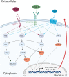
Major cellular pathways converge to promote angiogenic processes. The cellular receptors PDGFR, VEGFR-2, and CXCR-1, are activated upon ligand-binding and mediates activation of major signaling pathways such as MAPK/ERK, PI3K/Akt, PLCγ/PKC, and JAK/STAT. The activation of these pathways results in the transcription of pro-angiogenic genes that can promote cell migration, invasion, and angiogenesis in autocrine and paracrine manners.
FIGURE 3
FIGURE 3
Epstein–Barr virus proteins and miRNAs promote angiogenesis by activating several cellular signaling pathways. The viral transmembrane proteins LMP1 and LMP2A promote the activation of several signaling pathways including MAPK/ERK, PI3K/Akt, PLCγ/PKC, and JAK/STAT. EBNA1 directly promotes activation of pro-angiogenic genes and transcription factors such as AP-1. The BART miRNA targets the negative regulator of PI3K/Akt, PTEN. Activation of these signaling pathways results in the promotion of pro-survival and pro-angiogenic factors.
FIGURE 4
FIGURE 4
Several KSHV proteins and miRNAs promote angiogenesis by activating cellular signaling pathways. The viral factors LANA, vFLIP, Kaposin B, vIRF-3, and the miRNAs miR-K12 induces activation of major transcription factors such as HIF-1α, NF-κB, AP-1, and STAT-3. Additionally, the viral proteins K1, K15, vGPCR, vIL-6, and vPK cooperate in the activation of multiple signaling pathways promoting expression of angiogenic factors resulting in stronger viral tumorigenesis.

References

    1. Ablanedo-Terrazas Y., Alvarado-de la Barrera C., Ormsby C. E., Ruiz-Cruz M., Reyes-Teran G. (2015). Intralesional bevacizumab in patients with human immunodeficiency virus-associated Kaposi′s sarcoma in the upper airway. Laryngoscope 125 E132–E137. 10.1002/lary.24988 - DOI - PubMed
    1. Aga M., Bentz G. L., Raffa S., Torrisi M. R., Kondo S., Wakisaka N., et al. (2014). Exosomal HIF1alpha supports invasive potential of nasopharyngeal carcinoma-associated LMP1-positive exosomes. Oncogene 33 4613–4622. 10.1038/onc.2014.66 - DOI - PMC - PubMed
    1. Ahmed E. H., Baiocchi R. A. (2016). Murine models of Epstein-Barr virus-associated Lymphomagenesis. ILAR J. 57 55–62. 10.1093/ilar/ilv074 - DOI - PMC - PubMed
    1. An F. Q., Folarin H. M., Compitello N., Roth J., Gerson S. L., McCrae K. R., et al. (2006). Long-term-infected telomerase-immortalized endothelial cells: a model for Kaposi’s sarcoma-associated herpesvirus latency in vitro and in vivo. J. Virol. 80 4833–4846. 10.1128/JVI.80.10.4833-4846.2006 - DOI - PMC - PubMed
    1. Anders P. M., Montgomery N. D., Montgomery S. A., Bhatt A. P., Dittmer D. P., Damania B. (2018). Human herpesvirus-encoded kinase induces B cell lymphomas in vivo. J. Clin. Invest. 128 2519–2534. 10.1172/JCI97053 - DOI - PMC - PubMed

LinkOut - more resources