. 2019 Jul 4:2019:4124865.
doi: 10.1155/2019/4124865. eCollection 2019.

Pathology of A(H5N8) (Clade 2.3.4.4) Virus in Experimentally Infected Chickens and Mice

Affiliations

Pathology of A(H5N8) (Clade 2.3.4.4) Virus in Experimentally Infected Chickens and Mice

Elena A Prokopyeva et al. Interdiscip Perspect Infect Dis. .

Abstract

The emergence of novel highly pathogenic avian influenza viruses (HPAIVs) in migratory birds raises serious concerns as these viruses have the potential to spread during fall migration. We report the identification of novel HPAIV A(H5N8) clade 2.3.4.4 virus that was isolated from sick domestic duck at commercial farm during the second wave of spread that began in October and affected poultry (ducks; chiсkens) in several European regions of Russia and Western Siberia in 2016. The strain was highly lethal in experimental infection of chickens and mice with IVPI = 2.34 and MLD50 = 1.3log10⁡ EID50, accordingly. Inoculation of chickens with the HPAIV A/H5N8 demonstrated neuroinvasiveness, multiorgan failure, and death of chickens on the 3rd day post inoculation. Virus replicated in all collected organ samples in high viral titers with the highest titer in the brain (6.75±0.1 log10TCID50/ml). Effective virus replication was found in the following cells: neurons and glial cells of a brain; alveolar cells and macrophages of lungs; epithelial cells of a small intestine; hepatocytes and Kupffer cells of a liver; macrophages and endothelial cells of a spleen; and the tubular epithelial cells of kidneys. These findings advance our understanding of histopathological effect of A(H5N8) HPAIV infection.


Figures

Figure 1
Figure 1
Mean titers of A/domestic duck/Siberia/49feather/2016(H5N8) virus in organs of chickens. Chickens were infected iv with 106  EID50/100 μl of A/domestic duck/Siberia/49feather/2016(H5N8) virus. Note: viral titers were determined in MDCK cell culture by Kerber method and expressed as log10⁡   TCID50 in 1 ml of studied sample as М±CI95, where M is an arithmetic mean value and CI is a confidence interval.
Figure 2
Figure 2
Histological lesions and immunohistochemical detection of viral antigen in lungs, brain, and liver of chicken infected with A(H5N8) HPAI virus; viral antigen staining in red. Note: (a) erythrocytes in the parabronchial lumen and atria of the lung. Bar = 50 μm. (b) The same. Bar = 20 μm. (c) Viral antigen in the alveolar macrophages and alveolar cells. Bar = 20 μm. (d) Venous and capillary hyperemia of the brain. Bar = 100 μm. (e) Local area of brain infiltration. Bar = 20 μm. (f) Many AIV antigens are found in neurons (insert with magnification x1000) and glial cells. Bar = 20 μm. (g) The venous and capillary hyperemia in liver. Bar = 20 μm. (h) The dystrophic changes at a vast area of liver parenchyma; the predominance of active macrophages; and a large number of mast cells, basophils, and eosinophils. Bar = 100 μm. (i) Viral antigen in the Kupffer cell (insert with magnification x1000) and in hepatocytes (bar = 20 μm). Insert demonstrates viral antigen presence in hepatocyte. Magnification x1000. HE: hematoxylin-and-eosin staining; IHC: immunohistochemical staining.
Figure 3
Figure 3
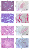
Histological lesions and immunohistochemical detection of viral antigen in heart, intestine, kidneys, and spleen of chicken infected with HPAI A(H5N8) virus; viral antigen staining in red. Note: (a) the lymphohistiocytic myocarditis. Bar = 20 μm. (b) Viral antigen in myocytes. Bar = 20 μm. (c) Intensive mononuclear infiltration of the small intestine; desquamation of epithelium. Bar = 20 μm. (d) Viral antigen in epithelial cells. Bar = 20 μm. E Venous and capillary hyperemia of kidney. Bar = 20 μm. (f) Viral antigens in kidney tubular epithelial cells. Magnification x40. (g) Local area of necrosis in spleen. Bar = 20 μm. (h) Viral antigen in spleen macrophages. Bar = 20 μm. HE: hematoxylin-and-eosin staining; IHC: immunohistochemical staining.

References

    1. Wan X. F. Isolation and Characterization of Avian Influenza Viruses in China. Guangzhou, China: College of Veterinary Medicine, South China Agricultural University; 1998.
    1. Zhao K., Gu M., Zhong L., et al. Characterization of three H5N5 and one H5N8 highly pathogenic avian influenza viruses in China. Veterinary Microbiology. 2013;163:351–357. doi: 10.1016/j.vetmic.2012.12.025. - DOI - PubMed
    1. Jeong J., Kang H., Lee E., et al. Highly pathogenic avian influenza virus (H5N8) in domestic poultry and its relationship with migratory birds in South Korea during 2014. Veterinary Microbiology. 2014;173:249–257. doi: 10.1016/j.vetmic.2014.08.002. - DOI - PubMed
    1. Marchenko V. Y., Susloparov I. M., Kolosova N. P., et al. Influenza A(H5N8) virus isolation in Russia, 2014. Archives of Virology. 2015;160:2857–2860. doi: 10.1007/s00705-015-2570-4. - DOI - PubMed
    1. Lee D., Sharshov K., Swayne D. E., et al. Novel reassortant clade 2.3.4.4 avian influenza A(H5N8) virus in wild aquatic birds, russia, 2016. Emerging Infectious Diseases. 2017;23(2):359–360. doi: 10.3201/eid2302.161252. - DOI - PMC - PubMed

LinkOut - more resources