. 2019 Jul 4:2019:7975407.
doi: 10.1155/2019/7975407. eCollection 2019.

Neuroinflammation and Myelin Status in Alzheimer's Disease, Parkinson's Disease, and Normal Aging Brains: A Small Sample Study

Affiliations

Neuroinflammation and Myelin Status in Alzheimer's Disease, Parkinson's Disease, and Normal Aging Brains: A Small Sample Study

Fei Han et al. Parkinsons Dis. .

Abstract

Microglia and astrocytes play important roles in mediating the immune processes and nutritional support in the central nervous system (CNS). Neuroinflammation has been indicated in the progression of neurodegenerative diseases Alzheimer's disease (AD) and Parkinson's disease (PD). Chronic neuroinflammation with sustained activation of microglia and astrocytes may affect white matter tracts and disrupt communication between neurons. Recent studies indicate astrogliosis may inhibit remyelination in demyelinating disorders such as multiple sclerosis. In this study, we investigated the relationship between neuroinflammation and myelin status in postmortem human brain tissue (n = 15 including 6 AD, 5 PD, and 4 age-matched, neurologically normal controls (NC)). We conducted systematic and quantitative immunohistochemistry for glial fibrillary acidic protein (GFAP), ionized calcium-binding adaptor molecule 1 (Iba1), amyloid beta, and highly phosphorylated tau (tauopathy). White matter intactness was evaluated by myelin and axon staining in adjacent brain tissue sections. Eight of 15 cases (4 AD, 3 PD, and 1 NC) showed increased immunoreactivity for microglia and astrocytes in the white matter that connects striatum and cortex. Quantitative analysis of these 8 cases showed a significant negative correlation between GFAP (but not Iba-1) and myelin (but not axon) staining in white matter (r 2 = 0.78, p < 0.005). Tau, but not amyloid beta plaques, is significantly higher in AD vs. PD and NC. Tau burden increases with age in AD cases. These observations indicate that astrocytosis in white matter is associated with loss of myelin in AD, PD, and normal aging and that tau is a potent biomarker for AD.


Figures

Figure 1
Figure 1
Myelin staining in the representative AD, PD, and age-matched cognitive intact controls (NC) cases. (a, c, e) The connected fine fibers between the junction of subcortical white and gray matter from AD, PD, and NC, respectively (the bars represent a 200 μm scale; the insets show a gross view, and the small squares indicate the zoomed regions). (b, d, f) The fibers in the subcortical white matter from the same AD, PD, and NC cases, respectively. The fibers are short and small in diameter, they are arranged in complex cross-linking pattern (the bars represent a 200 μm scale).
Figure 2
Figure 2
Astrocytes and microglia in the striatal tissue sections using GFAP and Iba1 antibodies. (a) Representative images of activated astrocytes and microglia in white matter, gray matter, and striatum. The activated astrocytes showed enlarged cell bodies and enlongated fibers, while activated microglia showed meatball-like appearance with enlarged cell bodies and shortened processes. The scale bar represents 100 μm. (b, c) The quantitative analysis of the GFAP and Iba1 expression in white matter in the astrocytosis and the nonastrocytosis groups. The bars represent mean ± SEM. represents p < 0.05.
Figure 3
Figure 3
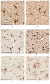
Different morphologies of microglia in AD, PD, and NC. (a, b) Dystrophic microglia from striatal white matter and cortex of two AD patients which displayed twisted or fragmented processes and cell body enlargement. (c, d) Activated and dystrophic microglia from striatum of two PD patients which showed cell body enlargement and fragmented processes. (e, f) Resting microglia from striatum of two health controls which showed the small cell body with highly branched processes. The scale represents 400 μm.
Figure 4
Figure 4
GFAP, Iba1, myelin, and axon staining and correlation analysis between GFAP expression and myelin density. Left panel shows the representative IHC images of GFAP, Iba1, myelin, and axon staining in the striatal tissue sections from AD, PD, and NC cases in the astrocytosis (a) and the nonastrocytosis group (c). Right panel shows the correlation between GFAP expression and myelin density in the astrocytosis (b) and the nonastrocytosis (d) groups.
Figure 5
Figure 5
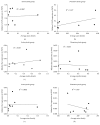
Correlation analysis between GFAP and Iba1 staining with axon and myelin densities. (a, b) The correlation between the expression of GFAP and axon densities in the astrocytosis and nonastrocytosis group. (c, d) The correlation between the expression of Iba1 and myelin densities in the astrocytosis and nonastrocytosis group. (e, f) The correlation between the expression of Iba1 and axon densities in the astrocytosis and nonastrocytosis group.
Figure 6
Figure 6
Relative GFAP and Iba1 expression and myelin and axon status in PD, AD, and age-matched cognitive intact controls (NC). (a) Expression of GFAP in the striatum. (b) Expression of GFAP in white matter. (c) Expression of GFAP in gray matter. (d) Expression of Iba1 in the striatum. (e) Expression of Iba1 in white matter. (f) Expression of Iba1 in gray matter. (g) Myelin status in AD, PD, and NC. (h) Axon status in AD, PD, and NC. The bars represent mean ± SEM.
Figure 7
Figure 7
Abnormal tau expression in AD patients. (a) Neurofibrillary tangle deposition in a 75-year-old male patient. (b) Neurofibrillary tangle deposition in a 93-year-old male patient (insets represent gross image, the small squares indicate the zoomed regions, and the bars represent a 200 μm scale). (c) Quantitative analysis of tau expression in six AD cases. (d) Quantitative analysis of tau and Aβ expression in the gray matter of six AD cases.
Figure 8
Figure 8
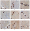
Immunohistochemical staining of α-SMA in AD, PD, and age-matched cognitively normal controls (NC) cases. The positive staining (brown color) were localized in the perivascular area (pericytes). The nucleus was stained by hematoxylin. The bars represent a 100 μm scale. (a) AD1, (b) AD2, (c) AD3, (d) PD1, (e) PD2, (f) PD3, (g) NC1, (h) NC2, (i) NC3.

References

    1. Bozzali M., Falini A., Franceschi M., et al. White matter damage in Alzheimer’s disease assessed in vivo using diffusion tensor magnetic resonance imaging. Journal of Neurology, Neurosurgery & Psychiatry. 2002;72(6):742–746. doi: 10.1136/jnnp.72.6.742. - DOI - PMC - PubMed
    1. Jackson J., Bianco G., Rosa A. O., et al. White matter tauopathy: transient functional loss and novel myelin remodeling. Glia. 2018;66(4):813–827. doi: 10.1002/glia.23286. - DOI - PubMed
    1. Mancardi G., Hart B., Roccatagliata L., et al. Demyelination and axonal damage in a non-human primate model of multiple sclerosis. Journal of the Neurological Sciences. 2001;184(1):41–49. doi: 10.1016/s0022-510x(00)00490-1. - DOI - PubMed
    1. Grueter B. E., Schulz U. G. Age-related cerebral white matter disease (leukoaraiosis): a review. Postgraduate Medical Journal. 2012;88(1036):79–87. doi: 10.1136/postgradmedj-2011-130307. - DOI - PubMed
    1. Phillips O. R., Clark K. A., Luders E., et al. Superficial white matter: effects of age, sex, and hemisphere. Brain Connectivity. 2013;3(2):146–159. doi: 10.1089/brain.2012.0111. - DOI - PMC - PubMed

LinkOut - more resources