Case Reports
. 2019 Jul 2:2019:8132578.
doi: 10.1155/2019/8132578. eCollection 2019.

Peripherally Embolizing Aortic Thrombus: The Work-Up, Management, and Outcome of Primary Aortic Thrombus

Affiliations
Case Reports

Peripherally Embolizing Aortic Thrombus: The Work-Up, Management, and Outcome of Primary Aortic Thrombus

Ramy Mando et al. Case Rep Cardiol. .

Abstract

Background: Primary aortic thrombus is an uncommon entity and not frequently reported in the literature. Herein, we discuss the presentation and management of a patient with a primary thoracic mural thrombus.

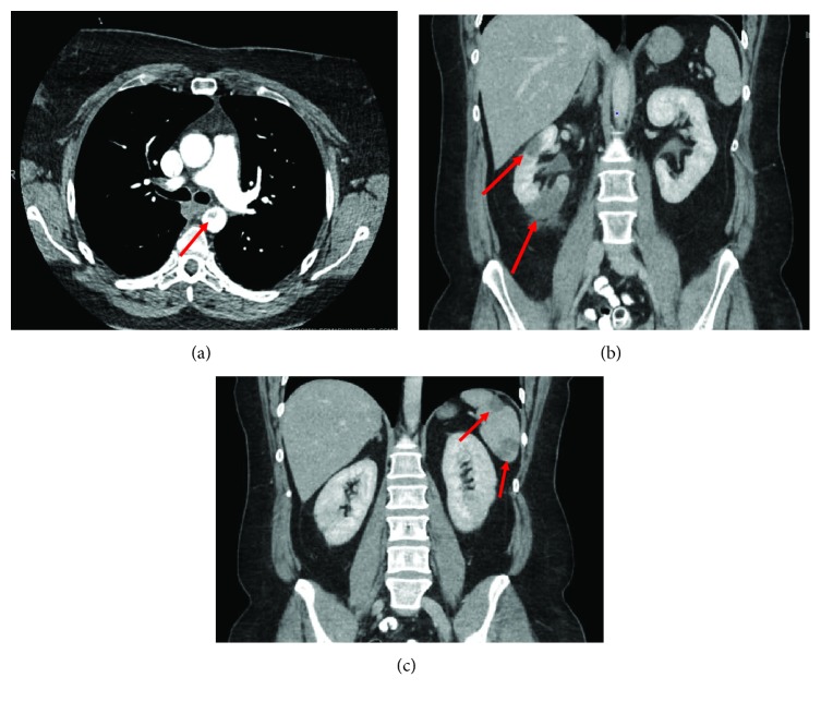
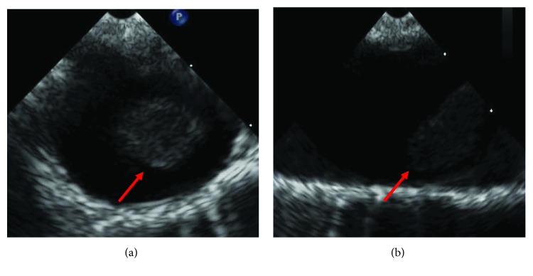
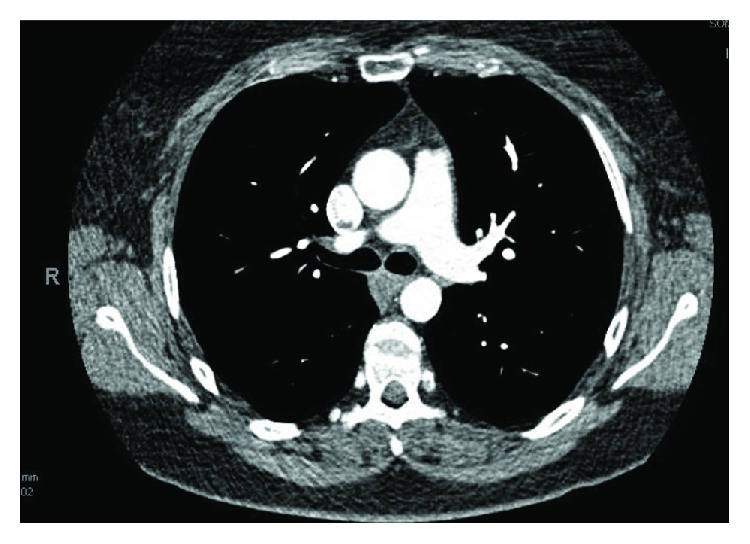
Case summary: A 46-year-old female with past medical history of tobacco dependence presented for low-grade fever and sudden onset, severe right upper quadrant abdominal pain with associated nausea and vomiting. Computed tomography (CT) revealed an intraluminal polypoid filling defect arising from the isthmus of the aorta projecting into the proximal descending aorta and findings consistent with infarction of the spleen and right kidney. Infectious, autoimmune, hematologic, and oncologic work-up were all unyielding. The patient was started on heparin and later transitioned to apixaban 5 mg twice a day and 81 mg of aspirin daily. She was also counseled regarding smoking cessation. Two months follow-up CT revealed resolution of the thrombus. Patient had no further thromboembolic complications.

Discussion: We present a unique case of primary aortic thrombus. To our knowledge, this is the first reported case managed successfully with a NOAC. This diagnosis is one of exclusion and through work-up should be completed. Our aim is to raise awareness of this condition and successful management with apixaban in low-risk patients.


Figures

Figure 1
Figure 1
CT of the abdomen and pelvis revealing a mass in the descending thoracic aortia (a) with wedge-shaped infarctions in the right kidney (b) and spleen (c) consistent with an embolic phenomona.
Figure 2
Figure 2
Apical four-chamber view of the heart with no evidence of a source of thrombosis.
Figure 3
Figure 3
Agitated saline (highlighted in red) in the right heart revealing no crossover into the left heart consistent with no intracardiac shunts.
Figure 4
Figure 4
(a) Transesophageal echocardiogram (transverse view) of a large mass attached to the wall of the descending thoracic aorta. (b) Transesophageal echocardiogram (longitudinal view) of the large mobile mass attached to the aortic wall.
Figure 5
Figure 5
Lower extremity arterial Doppler with significant flow limitations in the left lower extremity consistent with diffuse obstructive atherosclerotic obstructive disease in the left posterior tibial artery, peroneal artery, and anterior tibial artery. The waveforms of the right lower extremity are normal multiphasic Doppler waveforms. In the left extremity, we see dampened monophasic Doppler flow signals in the area of the left posterior tibial artery and dorsalis pedis artery. Diminished pulse volume recordings were seen in the left ankle. Flat line, nonpulsatile flow signals were seen in the digits of the left foot.
Figure 6
Figure 6
CT obtained for follow-up which reveals resolution of previously noted thrombus of the thoracic aorta.

Similar articles

Cited by

References

    1. Laperche T., Laurian C., Roudaut R., Steg P. G. Mobile thromboses of the aortic arch without aortic debris. A transesophageal echocardiographic finding associated with unexplained arterial embolism. Circulation. 1997;96(1):288–294. doi: 10.1161/01.CIR.96.1.288. - DOI - PubMed
    1. Tsilimparis N., Hanack U., Pisimisis G., Yousefi S., Wintzer C., Ruckert R. I. Thrombus in the non-aneurysmal, non-atherosclerotic descending thoracic aorta—an unusual source of arterial embolism. European Journal of Vascular and Endovascular Surgery. 2011;41(4):450–457. doi: 10.1016/j.ejvs.2010.11.004. - DOI - PubMed
    1. Verma H., Meda N., Vora S., George R. K., Tripathi R. K. Contemporary management of symptomatic primary aortic mural thrombus. Journal of Vascular Surgery. 2014;60(6):1524–1534. doi: 10.1016/j.jvs.2014.08.057. - DOI - PubMed
    1. Morris M. E., Galinanes E. L., Nichols W. K., Ross C. B., Chauvupun J. Thoracic mural thrombi: a case series and literature review. Annals of Vascular Surgery. 2011;25(8):1140.e17–1140.e21. doi: 10.1016/j.avsg.2011.05.030. - DOI - PubMed
    1. Fayad Z. Y., Semaan E., Fahoum B., Briggs M., Tortolani A., D’Ayala M. Aortic mural thrombus in the normal or minimally atherosclerotic aorta. Annals of Vascular Surgery. 2013;27(3):282–290. doi: 10.1016/j.avsg.2012.03.011. - DOI - PubMed

Publication types

LinkOut - more resources