

A Fatal Case of Large Cell Neuroendocrine Lung Cancer Metastatic to the Brain: A Case Report
- PMID: 31355087
- PMCID: PMC6649918
- DOI: 10.7759/cureus.4728
A Fatal Case of Large Cell Neuroendocrine Lung Cancer Metastatic to the Brain: A Case Report
Abstract
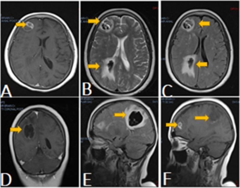
Large cell neuroendocrine carcinoma (LCNEC) is part of lung neuroendocrine tumors. LCNEC represents an extremely rare entity with aggressive behavior and poor prognosis. Primary surgery is the mainstream of treatment, although it is rarely amenable due to local or systemic tumor metastasis at the time of the diagnosis. We present a case report of a female patient diagnosed with large cell neuroendocrine lung cancer metastatic to the brain. Noting the low incidence of the disease, the lack of relevant clinical data has resulted in a challenge in diagnosis and management.
Keywords: brain metastasis; large cell neuroendocrine carcinoma; palliative radiation.
Conflict of interest statement
The authors have declared that no competing interests exist.
Figures


Similar articles
-
Breast metastasis and lung large-cell neuroendocrine carcinoma: first clinical observation.Clin Respir J. 2017 Sep;11(5):574-578. doi: 10.1111/crj.12385. Epub 2015 Oct 20. Clin Respir J. 2017. PMID: 26365150
-
Laparoscopic hepatectomy for liver metastasis of lung large-cell neuroendocrine carcinoma: A case report.Int J Surg Case Rep. 2019;65:40-43. doi: 10.1016/j.ijscr.2019.10.026. Epub 2019 Oct 21. Int J Surg Case Rep. 2019. PMID: 31678698 Free PMC article.
-
Gamma Knife radiosurgery for brain metastases from pulmonary large cell neuroendocrine carcinoma: a Japanese multi-institutional cooperative study (JLGK1401).J Neurosurg. 2016 Dec;125(Suppl 1):11-17. doi: 10.3171/2016.7.GKS161459. J Neurosurg. 2016. PMID: 27903179
-
Large Cell Neuroendocrine Carcinoma of the Prostate: A Systematic Review and Pooled Analysis.Urol Int. 2019;103(4):383-390. doi: 10.1159/000499883. Epub 2019 Apr 9. Urol Int. 2019. PMID: 30965328
-
Treatment of lung large cell neuroendocrine carcinoma.Tumour Biol. 2016 Jun;37(6):7047-57. doi: 10.1007/s13277-016-5003-4. Epub 2016 Mar 4. Tumour Biol. 2016. PMID: 26943800 Review.
Cited by
-
Metastatic large cell neuroendocrine lung cancer to the foramen magnum: A case report.Medicine (Baltimore). 2020 Aug 14;99(33):e21628. doi: 10.1097/MD.0000000000021628. Medicine (Baltimore). 2020. PMID: 32872023 Free PMC article.
-
Management of an Unusual Central Nervous System Metastasis With Linear Accelerator Radiosurgery in a Low-Middle Income Country.Cureus. 2021 Nov 22;13(11):e19806. doi: 10.7759/cureus.19806. eCollection 2021 Nov. Cureus. 2021. PMID: 34956790 Free PMC article.
-
Large cell neuroendocrine tumor of lung with hemorrhagic cerebral metastasis: A case report.Radiol Case Rep. 2025 Jun 26;20(9):4679-4689. doi: 10.1016/j.radcr.2025.06.002. eCollection 2025 Sep. Radiol Case Rep. 2025. PMID: 40677880 Free PMC article.
References
-
- Treatment outcomes and incidence of brain metastases in pulmonary large cell neuroendocrine carcinoma. Zhao Y, Castonguay M, Wilke D, et al. Curr Prob Cancer. 2019;43:54–65. - PubMed
Publication types
LinkOut - more resources
Full Text Sources