. 2019 Jul 10:9:619.
doi: 10.3389/fonc.2019.00619. eCollection 2019.

MicroRNA-153 Decreases Tryptophan Catabolism and Inhibits Angiogenesis in Bladder Cancer by Targeting Indoleamine 2,3-Dioxygenase 1

Affiliations

MicroRNA-153 Decreases Tryptophan Catabolism and Inhibits Angiogenesis in Bladder Cancer by Targeting Indoleamine 2,3-Dioxygenase 1

Wentao Zhang et al. Front Oncol. .

Erratum in

Abstract

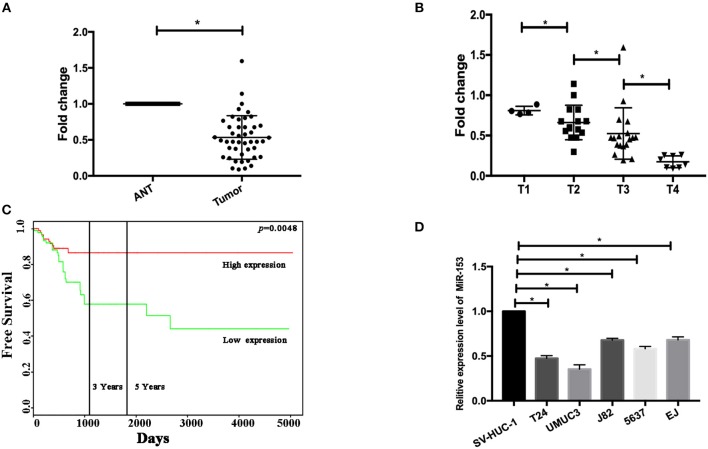
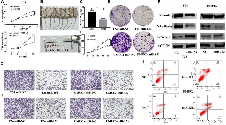
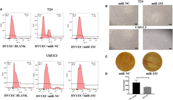
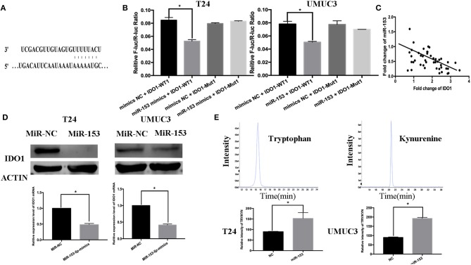
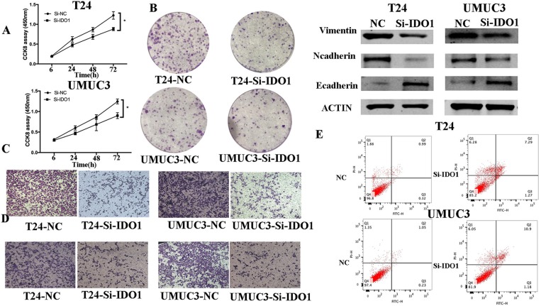
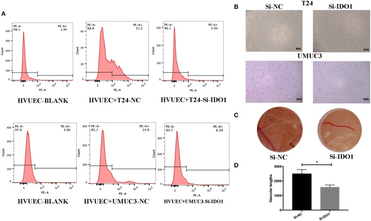
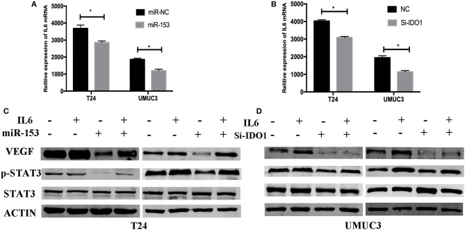
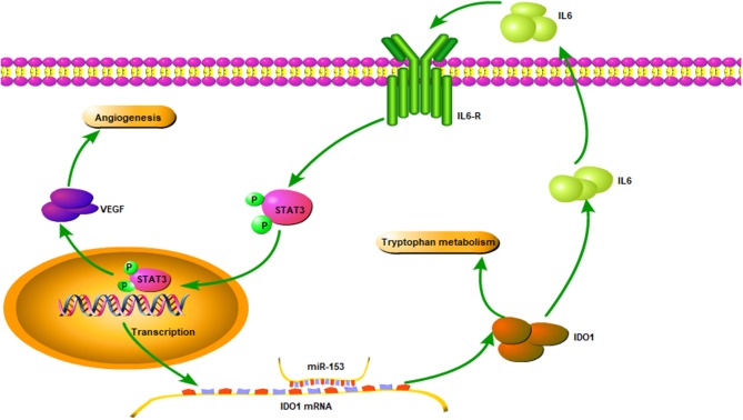
Background: Metastasis is the primary cause of cancer deaths, warranting further investigation. This study assessed microRNA-153 (miR-153) expression in bladder cancer tissues and investigated the underlying molecular mechanism of miR-153-mediated regulation of bladder cancer cells. Methods: Paired tissue specimens from 45 bladder cancer patients were collected for qRT-PCR. The Cancer Genome Atlas (TCGA) dataset was used to identify associations of miR-153 with bladder cancer prognosis. Bladder cancer tissues and immortalized cell lines were used for the following experiments: miR-153 mimics and indoleamine 2,3-dioxygenase 1 (IDO1) siRNA transfection; Western blot, cell viability, colony formation, and Transwell analyses; nude mouse xenograft; and chicken embryo chorioallantoic membrane angiogenesis (CAM) assays. Human umbilical vein endothelial cells (HUVECs) were co-cultured with bladder cancer cells for the tube formation assay. The luciferase reporter assay was used to confirm miR-153-targeting genes. Results: miR-153 expression was downregulated in bladder cancer tissues and cell lines, and reduced miR-153 expression was associated with advanced tumor stage and poor overall survival of patients. Moreover, miR-153 expression inhibited bladder cancer cell growth by promoting tumor cell apoptosis, migration, invasion, and endothelial mesenchymal transition (EMT) in vitro and tumor xenograft growth in vivo, while miR-153 expression suppressed HUVEC and CAM angiogenesis. At the gene level, miR-153 targeted IDO1 expression and inhibited bladder cancer cell tryptophan metabolism through inhibiting IL6/STAT3/VEGF signaling. Conclusions: Collectively, our data demonstrate that miR-153 exerts anti-tumor activity in bladder cancer by targeting IDO1 expression. Future studies will investigate miR-153 as a novel therapeutic target for bladder cancer patients.

Keywords: 3-dioxygenase 1; angiogenesis; bladder cancer; indoleamine 2; miR-153; tryptophan catabolism.


Figures

Figure 1
Figure 1
miR-153 is downregulated in bladder cancer tissues and cell lines. (A) qRT-PCR. miR-153 was detected in 45 pairs of bladder cancer (tumor) and adjacent normal tissues using qPCR (P < 0.05). (B) Association of miR-153 with clinicopathological features. Decreased miR-153 levels are associated with advanced tumor T stages. (C) Kaplan-Meier curve and log rank test stratified by miR-153 expression in TCGA dataset (http://tcga-data.nci.nih.gov/tcga/). (D) qRT-PCR. Various bladder cancer cell lines (T24, UMUC3, J82, 5637, and EJ) and an immortalized bladder epithelial cell line (SV-HUC-1) were grown and used for qPCR analysis (*P < 0.05).
Figure 2
Figure 2
miR-153 inhibits bladder cancer growth in vitro and in vivo by promoting tumor cell apoptosis, migration, invasion, and EMT. (A) Cell viability CCK-8 assay. T24 and UMUC3 cells were transfected with miR-153 mimics or negative control and then subjected to the CCK-8 assay. (B) Nude mouse xenograft assay. Stably miR-153 expressing mimics or negative control bladder cancer cells were subcutaneously injected into nude mice and monitored for 40 days for tumor cell xenograft formation and growth. (C) Tumor cell xenograft growth curves. (D) Tumor cell xenograft weight. (E) Colony formation assay. T24 and UMUC3 cells were transfected with miR-153 mimics or negative control and then subjected to tumor cell colony formation assay (x 200). (F) Western blot. Expression levels of EMT-associated markers in miR-153 mimics or negative control transfected T24 and UMUC3 cells were evaluated by using Western blot analysis. (G) Transwell tumor cell migration assay. (H) Transwell tumor cell invasion assay. (I) Flow cytometric Annexin V-PI double staining assay. *P < 0.05.
Figure 3
Figure 3
miR-153 inhibits bladder cancer angiogenesis in vitro and in vivo. (A) Flow cytometric assay. CD34 expression on HVUECs was assessed using flow cytometry in after co-culture with miR-153 or negative control transfected T24 and UMUC3 cells. (B) HUVEC tube formation assay. HUVECs were co-cultured with T24 and UMUC3 cells using Transwells and then subjected to the capillary-like tube formation assay (x100). (C) The CAM angiogenesis assay. miR-153 or negative control transfected T24 cells were added on to CAM to induce angiogenesis for 48 h to induce formation of vascular branches (x 10). (D) The vascular length are measured (*p < 0.05).
Figure 4
Figure 4
IDO1 is a direct miR-153 target. (A) Bioinformatical analysis predicted that IDO1 is a direct miR-153 target. (B) Luciferase reporter assay. miR-153 or miR-NC-transfected bladder cancer cells were co-transfected with a luciferase reporter plasmid (WT or MUT 3 -UTR IDO1 cDNA) and then subjected to protein extraction and luciferase reporter assay. (C) miR-153 is negatively correlated with the expression of IDO1 in cancer tissues by qRT-PCR (D) qRT-PCR and Western blot. miR-153 or negative control transfected T24 and UMUC3 cells were grown and subjected to qRT-PCR and Western blot. (E) UHPLC-MS. miR-153 or negative control transfected T24 and UMUC3 cells were grown and the supernatants were subjected to UHPLC-MS; comparison of the tryptophan ratio to kynurenine. *P < 0.05.
Figure 5
Figure 5
IDO1 knockdown inhibits bladder cancer cell proliferation, migration, and invasion, and induced apoptosis and modulation of EMT markers. (A) Cell viability CCK-8assay. T24 and UMUC3 cells were transfected with IDO1 or negative control siRNA and then subjected to the CCK-8 assay. (B) Colony formation assay. T24 and UMUC3 cells were transfected with IDO1 or negative control siRNA and then subjected to colony formation (x 200) and Transwell assays. (C) Western blot. Levels of the EMT-associated markers were analyzed in T24 and UMUC3 cells after IDO1 knockdown by using Western blot. (D) Transwell migration assay. (E) Transwell invasion assay. (E) Flow cytometric Annexin V-PI double staining assay in T24 and UMUC3 cells after knockdown of IDO1. *P < 0.05.
Figure 6
Figure 6
IDO1 knockdown inhibits angiogenesis. (A) Flow cytometric assay. HUVECs were co-cultured with IDO-siRNA or negative control siRNA-transfected T24 and UMUC3 cells and then subjected to flow cytometric analysis of CD34 level on HUVECs. (B) HUVEC tube formation assay. The same cultured HUVECs were subjected to the tube formation assay (x100). (C,D) The CAM assays. The siRNA-transfected T24 cells were added onto CAM to modulate angiogenesis for 48 h (x 10). The branches and length are measured (*p < 0.05).
Figure 7
Figure 7
miR-153 targets IDO1 and modulates angiogenesis through IL6/STAT3/VEGF signaling. (A,B) ELISA. IL_6 expression in T24 and UMUC3 cells (overexpression of miR-153 or knockdown of IDO1 and their respective negative controls) were analyzed using ELISA. (C,D) Western blot. T24 and UMUC3 cells (overexpression of miR-153 or knockdown of IDO1 and their respective negative controls) were pretreated with 100 ng/ml of IL-6 for 48 h and then subjected to Western blot analysis of STAT3, p-STAT3, and VEGF.
Figure 8
Figure 8
Illustration of the miR-153 gene pathway in bladder cancer cells. miR-153 expression is lost in bladder cancer, which decreases IDO1 expression and subsequent activity of the IL6/STAT3/VEGF signaling pathway in bladder cancer cells.

References

    1. Torre L, Bray F, Siegel R, Ferlay J, Lortet-Tieulent J, Jemal AJCCJC. Global cancer statistics, 2012. CA Cancer J Clin. (2015) 65:87–108. 10.3322/caac.21262 - DOI - PubMed
    1. Osch FHMV, Jochems SHJ, Schooten FJV, Bryan RT, Zeegers MP. Quantified relations between exposure to tobacco smoking and bladder cancer risk: a meta-analysis of 89 observational studies. Int J Epidemiol. (2016) 45:857. 10.1093/ije/dyw044 - DOI - PubMed
    1. Reulen RC, Kellen E, Buntinx F, Brinkman M, Zeegers MP. A meta-analysis on the association between bladder cancer and occupation. Scand J Urol Nephrol Suppl. (2008) 42:64–78. 10.1080/03008880802325192 - DOI - PubMed
    1. Alfred Witjes J, Lebret T, Comperat EM, Cowan NC, De Santis M, Bruins HM, et al. Updated 2016 EAU guidelines on muscle-invasive and metastatic bladder cancer. Eur Urol. (2017) 71:462–75 10.1016/j.eururo.2016.06.020 - DOI - PubMed
    1. Alexandroff AB, Jackson AM, O'Donnell MA, James K. BCG immunotherapy of bladder cancer: 20 years on. Lancet. (1999) 353:1689. 10.1016/S0140-6736(98)07422-4 - DOI - PubMed