. 2019 Jul 12:9:244.
doi: 10.3389/fcimb.2019.00244. eCollection 2019.

Extreme Diversity of Mycoviruses Present in Isolates of Rhizoctonia solani AG2-2 LP From Zoysia japonica From Brazil

Affiliations

Extreme Diversity of Mycoviruses Present in Isolates of Rhizoctonia solani AG2-2 LP From Zoysia japonica From Brazil

Maria Aurea S C Picarelli et al. Front Cell Infect Microbiol. .

Abstract

Zoysia japonica, in Brazil, is commonly infected by Rhizoctonia solani (R. solani) in humid and cool weather conditions. Eight isolates of R. solani, previously identified as belonging to the AG2-2 LP anastomosis group, isolated from samples from large path symptoms, were collected from three counties in São Paulo state (Brazil) and investigated for the presence of mycoviruses. After detection of double-strand RNA (dsRNA) in all samples, RNA_Seq analysis of ribosomal RNA-depleted total RNA from in vitro cultivated mycelia was performed. Forty-seven partial or complete viral unique RNA dependent-RNA polymerase (RdRp) sequences were obtained with a high prevalence of positive sense ssRNA viruses. Sequences were sufficiently different from the first match in BLAST searches suggesting that they all qualify as possible new viral species, except for one sequence showing an almost complete match with Rhizoctonia solani dsRNA virus 2, an alphapartitivirus. Surprisingly four large contigs of putative viral RNA could not be assigned to any existing clade of viruses present in the databases, but no DNA was detected corresponding to these fragments confirming their viral replicative nature. This is the first report on the occurrence of mycoviruses in R. solani AG2-2 LP in South America.

Keywords: Rhizoctonia solani; grass; multiple infection; mycoviruses; phylogenetic analysis; viral diversity; virus taxonomy.


Figures
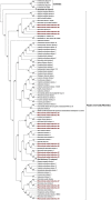
Figure 1
Figure 1
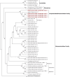
Phylogenetic analysis of positive sense RNA viruses related to the genus Mitovirus. 142 sequences have been used to produce an alignment starting from viruses belonging to the family Narnaviridae, Botourmiaviridae, and Leviviridae as outgroup; the phylogenetic tree was built using the maximum likelihood method, the best choice for the substitution model according to ModelFinder was PMB+F+I+G4. Ultrafast bootstrap analysis was performed with 1,000 replicates, and branches displaying values below 50 were collapsed. Viruses discovered in this work are in bold red ink; 42 sequences belonging to the Narnaviridae family have been compressed in one branch. A list of accession numbers of the sequences used for this analysis can be found in Supplementary Table 3, online. Detailed information about softwares used for the analysis can be found in the material and methods section.
Figure 2
Figure 2
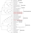
Phylogenetic analysis of positive sense RNA viruses related to the genus Botourmiaviridae. 142 sequences have been used to produce an alignment starting from viruses belonging to the family Narnaviridae, Botourmiaviridae, and Leviviridae as outgroup; phylogenetic tree was built using the maximum likelihood method the best choice for the substitution model according to ModelFinder was PMB+F+I+G4. Ultrafast bootstrap analysis was performed with 1,000 replicates, and branches displaying values below 50 were collapsed. Viruses discovered in this work are in bold red ink; 96 sequences belonging to the genus Mitovirus have been compressed in one branch. A list of accession numbers of the sequences used for this analysis can be found in Supplementary Table 3, online. Detailed information about softwares used for the analysis can be found in the material and methods section.
Figure 3
Figure 3
Phylogenetic analysis of viruses belonging to the family Endornaviridae. Twenty six viral sequences were used to build the alignment. A phylogenetic tree was built using the maximum likelihood method and the best substitution model selected by ModelFinder was VT+F+I+G4. Ultrafast bootstrap analysis was performed with 1,000 replicates and branches displaying values below 50 were collapsed. Viruses discovered in this work are outlined in bold red. Barley yellow mosaic virus, belonging to the family Potyviridae, was used as outgroup. A list of accession numbers of the sequences used for this analysis can be found in Supplementary Table 4, online. Detailed information about softwares used for the analysis can be found in the material and methods section.
Figure 4
Figure 4
Phylogenetic analysis of viruses belonging to the family Benyviridae. Sixteen viral sequences were aligned and a phylogenetic tree was derived using the maximum likelihood method. The best substitution model selected by ModelFinder was VT+F+I+G4. Ultrafast bootstrap analysis was performed with 1,000 replicates, and branches displaying values below 50 were collapsed. Viruses discovered in this work are outlined in bold red. Bromoviridae and Virgaviridae were used as outgroups. A list of accession numbers of the sequences used for this analysis can be found in Supplementary Table 4, online. Detailed information about softwares used for the analysis can be found in the material and methods section.
Figure 5
Figure 5
Phylogenetic analysis of viruses related to the Alphavirus supergroup. Forty two sequences were aligned and a phylogenetic tree was built using the maximum likelihood method. The best substitution model selected by ModelFinder was VT+F+I+G4 and ultrafast bootstrap analysis was performed with 1,000 replicates. Branches displaying values below 50 were collapsed. Viruses discovered in this work are outlined in bold red ink. A list of accession numbers of the sequences used for this analysis can be found in Supplementary Table 4, online. Detailed information about softwares used for the analysis can be found in the material and methods section.
Figure 6
Figure 6
Phylogenetic analysis of viruses related to the family Hypoviridae. Twenty eight sequences were used to build an alignment and to derive a phylogenetic tree using the maximum likelihood method. The best substitution model selected by ModelFinder was VT+F+G4 and an ultrafast bootstrap analysis was performed with 1,000 replicates. Viruses discovered in this work are outlined in bold red. Plum pox virus, belonging to the family Potyviridae, was used as outgroup. A list of accession numbers of the sequences used for this analysis can be found in Supplementary Table 5, online. Detailed information about softwares used for the analysis can be found in the material and methods section.
Figure 7
Figure 7
Phylogenetic analysis of viruses related to the orders Bunyavirales and Serpentovirales. Thirty seven sequences were aligned and the best substitution model selected by ModelFinder was VT+F+G4 and implemented in a maximum likelihood method to derive the Phylogenetic tree. Ultrafast bootstrap analysis was performed with 1,000 replicates and branches displaying values below 50 were collapsed. The Virus discovered in this work is outlined in red. A list of accession numbers of the sequences used for this analysis can be found in Supplementary Table 6, online. Detailed information about software used for the analysis can be found in the material and methods section.
Figure 8
Figure 8
Phylogenetic analysis of dsRNA viruses. Eighty four sequences were used to build an alignment. Phylogenetic tree was built using the maximum likelihood method, the best substitution model selected by ModelFinder was Blosum62+F+G4 and ultrafast bootstrap analysis was performed with 1,000 replicates, branches displaying values below 50 were collapsed. Viruses discovered in this work are underlined in red. A list of accession numbers of the sequences used for this analysis can be found in Supplementary Table 7, online. Detailed information about software used for the analysis can be found in the material and methods section.

References

    1. Andika I. B., Wei S., Cao C., Salaipeth L., Kondo H., Sun L. (2017). Phytopathogenic fungus hosts a plant virus: a naturally occurring cross-kingdom viral infection. Proc. Natl. Acad. Sci. U.S.A. 114, 12267–12272. 10.1073/pnas.1714916114 - DOI - PMC - PubMed
    1. Antoniolli D. (2015). “Produção, regularização e conquistas do mercado de gramas cultivadas no Brasil,” in Tópicos atuais em gramados IV, VII SIGRA – Simpósio sobre Gramados. FCA/UNESP. eds Mateus C.M.D'A., Villas Bôas R.L., Andrade T. F., Oliveira M.R., Backes C., Santos A.J.M., Godoy L.J.G. (Botucatu: Editora Fepaf, 9–22.
    1. Bartholomäus A., Wibberg D., Winkler A., Pühler A., Schlüter A., Varrelmann M. (2016). Deep sequencing analysis reveals the mycoviral diversity of the virome of an avirulent isolate of Rhizoctonia solani AG-2-2 IV. PLoS ONE 11:e0165965. 10.1371/journal.pone.0165965 - DOI - PMC - PubMed
    1. Bharathan N., Saso H., Gudipati L., Bharathan S., Whited K. (2005). Double-stranded RNA: distribution and analysis among isolates of Rhizoctonia solani AG-2 to−13. Plant Pathol. 54, 196–203. 10.1111/j.1365-3059.2005.01159.x - DOI
    1. Chiba S., Salaipeth L., Lin Y. H. (2009). A novel bipartite double-stranded RNA mycovirus from the white root rot fungus Rosellinia necatrix: molecular and biological characterization, taxonomic considerations, and potential for biological control. J. Virol. 3, 12801–12812. 10.1128/JVI.01830-09 - DOI - PMC - PubMed

MeSH terms