

Surface Electromyography of the Vastus Lateralis, Biceps Femoris, and Gluteus Medius in Dogs During Stance, Walking, Trotting, and Selected Therapeutic Exercises
- PMID: 31355214
- PMCID: PMC6636385
- DOI: 10.3389/fvets.2019.00211
Surface Electromyography of the Vastus Lateralis, Biceps Femoris, and Gluteus Medius in Dogs During Stance, Walking, Trotting, and Selected Therapeutic Exercises
Abstract
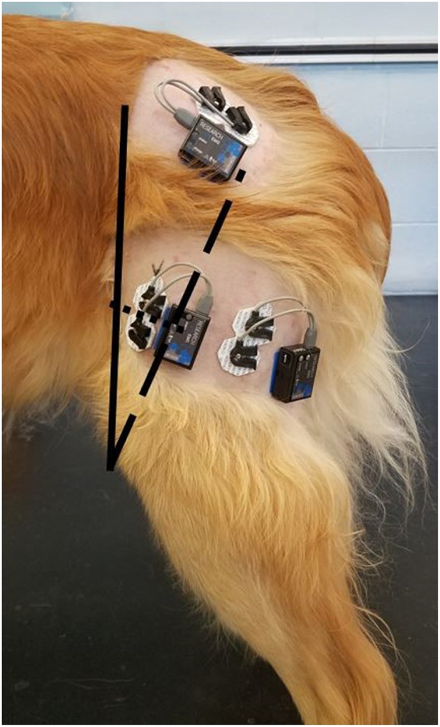
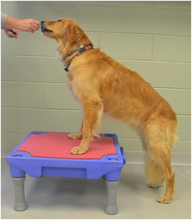
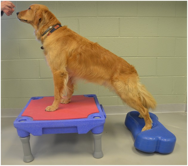
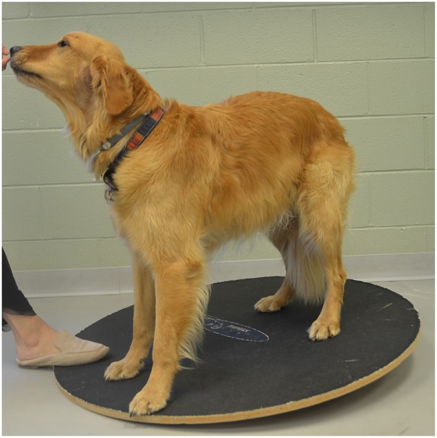
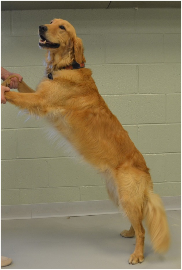
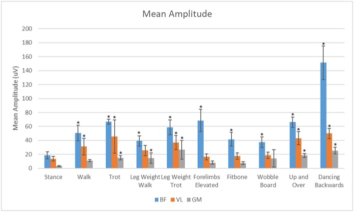
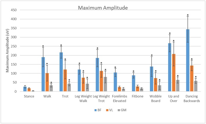
Objective: The objective of the study reported here was to evaluate the muscle activity patterns of the vastus lateralis (VL), biceps femoris (BF), and gluteus medius (GM) during stance, walking, trotting, and selected therapeutic exercises in clinically sound, healthy dogs. Our hypothesis was that the muscle activity during all exercises would differ from muscle activity at the stance. Methods: Surface electromyography of the selected muscles was performed during stance, walking, trotting, elevation of forelimbs on a platform, elevation of forelimbs on a platform with hindlimbs on an inflatable balance device, stepping up onto and over an obstacle, standing on a wobble board, dancing backwards, and wearing a leg weight at the walk and the trot. The maximal and mean muscle amplitude (μV) reflecting activity during several motion cycles were compared among the exercises. Results: Mean EMG amplitude of the BF was significantly higher in all exercises (p < 0.05) in comparison to stance. Mean EMG amplitude of the VL was significantly higher (p < 0.05) during walking, trotting, dancing backwards, stepping up and over an obstacle, and at a trot with a leg weight as compared to stance. Mean EMG amplitude of the GM was significantly higher (p < 0.05) during trotting, at a walk and a trot with a leg weight, standing on a wobble board, stepping up and over an obstacle, and dancing backwards when compared to stance. Of the studied exercises, dancing backwards increased the mean EMG amplitude of the BF and GM to the largest degree. Stepping up and over an obstacle increased the mean EMG amplitude of the VL to the largest degree. Conclusion: Compared to stance, the majority of therapeutic exercises examined increased muscle activity to varying degrees in the BF, VL, and GM. Our results may help clinicians to choose specific exercises to target specific muscles during conditioning, strengthening and rehabilitation.
Keywords: canine; exercise; physical therapy; rehabilitation; surface electromyography.
Figures

References
-
- Millis D, Levine D. Canine Rehabilitation and Physical Therapy. 2nd ed. Philadelphia, PA: Elsevier; (2014).
-
- Goslow G, Seeherman H, Taylor C, Mccutchin M, Heglund N. Electrical activity and relative length changes of dog limb muscles as a function of speed and gait. J Exp Biol. (1981) 94:15–42. - PubMed
LinkOut - more resources
Full Text Sources
Miscellaneous