

Glyceraldehyde-3-Phosphate Dehydrogenase of Babesia microti Is a Plasminogen- and Actin-Binding Protein
- PMID: 31355216
- PMCID: PMC6637311
- DOI: 10.3389/fvets.2019.00228
Glyceraldehyde-3-Phosphate Dehydrogenase of Babesia microti Is a Plasminogen- and Actin-Binding Protein
Abstract
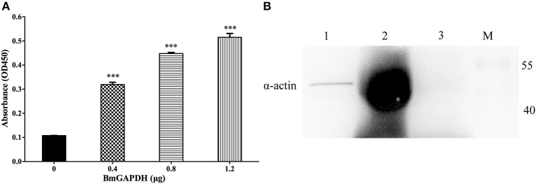
Babesia microti, an intraerythrocytic protozoa, can cause an emerging tick-borne disease-Human babesiosis. The parasite can successfully invade host red blood cells owing to the assistance of molecules expressed by babesia. Glyceraldehyde-3-phosphate dehydrogenase (GAPDH), the housekeeping intracellular glycolytic enzyme, can also be expressed in the external of cells, where contributes to binding to several molecules such as plasminogen and actin. In the present study, we identified B. microti GAPDH (BmGAPDH) and generated the recombinant BmGAPDH (rBmGAPDH) via an E. coli expression system. Furthermore, we confirmed its catalytic dehydration activity in vitro. Moreover, we also demonstrated that rBmGAPDH could bind to human plasminogen and mouse α-actin. In addition, we demonstrated that rBmGAPDH could recognize anti-B. microti mouse serum. In conclusion, BmGAPDH is a multifunctional glycolytic enzyme, which can bind to host plasminogen and α-actin.
Keywords: Babesia microti; binding protein; glyceraldehyde-3-phosphate dehydrogenase; plasminogen; α-actin.
Figures
Similar articles
-
Complete Molecular and Immunoprotective Characterization of Babesia microti Enolase.Front Microbiol. 2017 Apr 11;8:622. doi: 10.3389/fmicb.2017.00622. eCollection 2017. Front Microbiol. 2017. PMID: 28443086 Free PMC article.
-
Molecular characterization of Babesia microti thioredoxin (BmTrx2) and its expression patterns induced by antiprotozoal drugs.Parasit Vectors. 2018 Jan 15;11(1):38. doi: 10.1186/s13071-018-2619-9. Parasit Vectors. 2018. PMID: 29335000 Free PMC article.
-
Case report of the patient source of the Babesia microti R1 reference strain and implications for travelers.J Travel Med. 2018 Jan 1;25(1):tax073. doi: 10.1093/jtm/tax073. J Travel Med. 2018. PMID: 29394381 Free PMC article.
-
Vaccination against babesiosis using recombinant GPI-anchored proteins.Int J Parasitol. 2019 Feb;49(2):175-181. doi: 10.1016/j.ijpara.2018.12.002. Epub 2019 Jan 24. Int J Parasitol. 2019. PMID: 30684517 Review.
-
Splenic Rupture as the First Manifestation of Babesia Microti Infection: Report of a Case and Review of Literature.Am J Case Rep. 2018 Mar 23;19:335-341. doi: 10.12659/ajcr.908453. Am J Case Rep. 2018. PMID: 29567936 Free PMC article. Review.
Cited by
-
Molecular characterization of glyceraldehyde-3-phosphate dehydrogenase from Eimeria tenella.Parasitol Res. 2022 Jun;121(6):1749-1760. doi: 10.1007/s00436-022-07508-5. Epub 2022 Apr 2. Parasitol Res. 2022. PMID: 35366097
-
Proteomic analysis of Taenia solium cysticercus and adult stages.Front Vet Sci. 2023 Jan 9;9:934197. doi: 10.3389/fvets.2022.934197. eCollection 2022. Front Vet Sci. 2023. PMID: 36699330 Free PMC article.
-
Glyceraldehyde-3-phosphate dehydrogenase (GAPDH) moonlights as an adhesin in Mycoplasma hyorhinis adhesion to epithelial cells as well as a plasminogen receptor mediating extracellular matrix degradation.Vet Res. 2021 Jun 3;52(1):80. doi: 10.1186/s13567-021-00952-8. Vet Res. 2021. PMID: 34082810 Free PMC article.
References
LinkOut - more resources
Full Text Sources
Research Materials