Review
. 2019 Jun;7(12):269.
doi: 10.21037/atm.2019.05.37.

MR imaging for acute pancreatitis: the current status of clinical applications

Affiliations
Review

MR imaging for acute pancreatitis: the current status of clinical applications

Huan Sun et al. Ann Transl Med. 2019 Jun.

Abstract

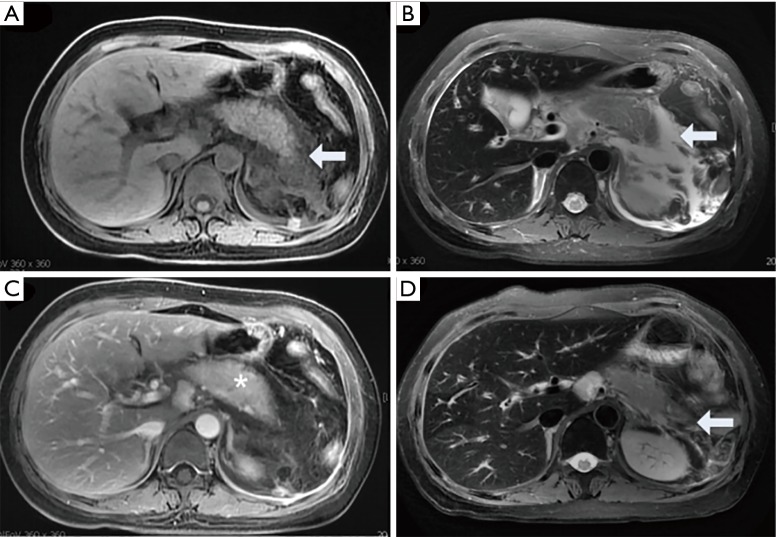
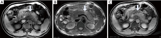
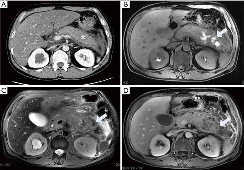
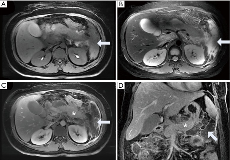
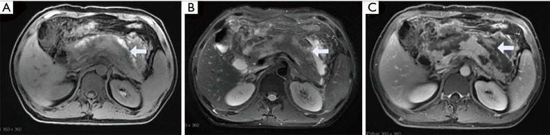
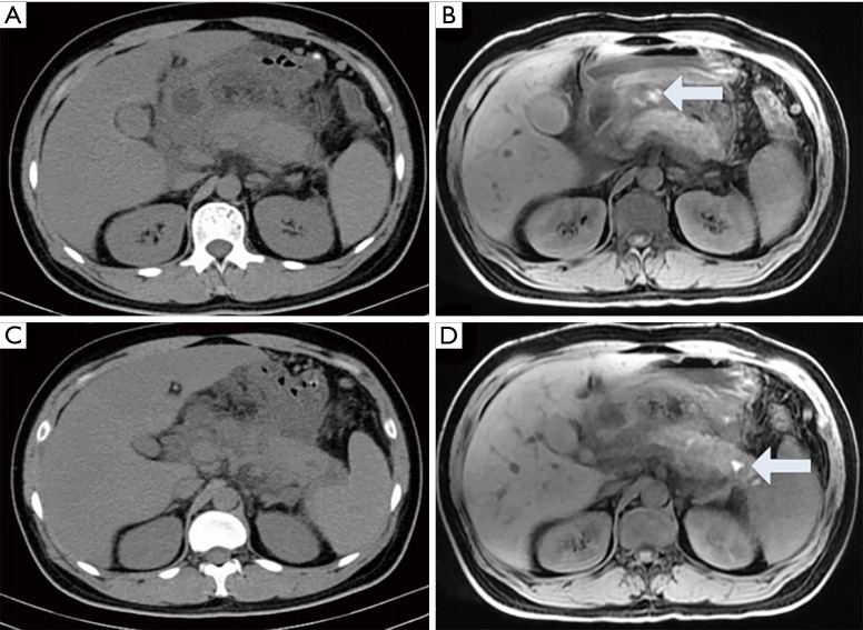
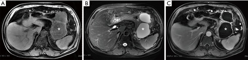
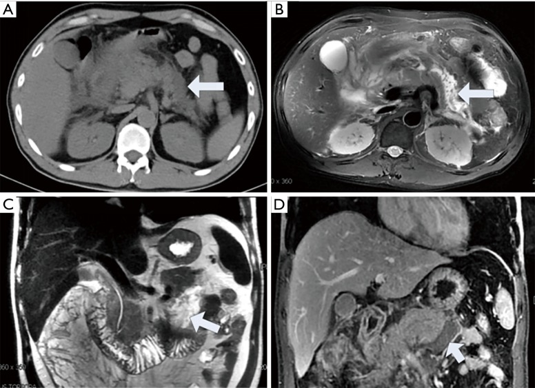
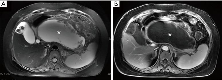
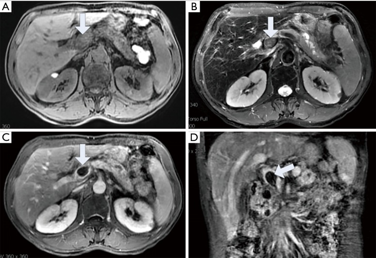
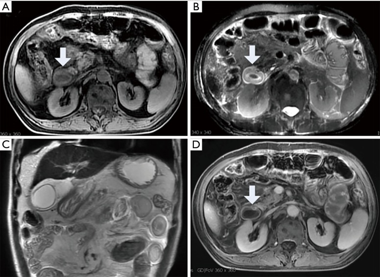
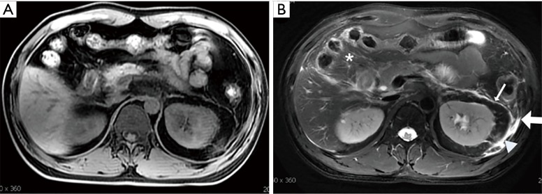
Acute pancreatitis is a common clinical acute abdomen. Imaging examinations play an important role in the management of acute pancreatitis. MR imaging is a noninvasive examination with high tissue contrast and a variety of acquisition sequences that can help determine the diagnosis, complications and severity of acute pancreatitis. The acute pancreatitis classification working group modified the Atlanta classification in 2012 to improve clinical evaluations and standardize the radiologic nomenclature for acute pancreatitis. In particular, the redefinition of necrotizing pancreatitis offers a new understanding of this disease. In clinical practice, there is still a lack of unifying standards between radiologists and physicians, such as for the imaging features of pseudocysts, walled-off necrosis, peripancreatic necrosis and especially for the MR imaging features of acute pancreatitis. In this article, we review the 2012 revised Atlanta classification of acute pancreatitis and recent advances in the clinical applications of MR imaging (MRI) in acute pancreatitis by showing how MRI can provide more optimized information for clinical diagnosis and treatment plan.

Keywords: MR imaging (MRI); acute pancreatitis (AP); local complication.


Conflict of interest statement

Conflicts of Interest: The authors have no conflicts of interest to declare.

Figures

Figure 1
Figure 1
A 61-year-old male with acute edematous pancreatitis. On the third day after onset, the pancreas showed swelling and there was homogeneous peripancreatic fluid collection on the axial T1WI (A, arrow) and the T2WI (B, arrow). The pancreas was homogeneous enhanced after contrast injection (C, asterisk). After 9 days of treatment, the peripancreatic collection was absorbed (D, arrow).
Figure 2
Figure 2
A 45-year-old male with acute necrotizing pancreatitis within 3 days after onset (pancreatic parenchymal necrosis alone). The neck and body of the pancreas show hypointensity combined with patchy isointensity (arrow) on the T1WI (A), high and low mixed signal intensity on the T2WI (B, arrow), and no enhancement after an intravenous injection of contrast material (C, arrow). The extent of necrosis is approximately 30–50% of the pancreatic gland. In the peripancreatic tissue, stripe-like hyperintensity is observed on the T2WI (B).
Figure 3
Figure 3
A 55-year-old male with acute necrotizing pancreatitis. On the first day after onset, the enhanced CT image (A) showed enlargement of the body and tail of the pancreas, collections of peripancreatic fluid, and homogeneous enhancement of the pancreatic parenchyma (asterisk). However, on the 9th day after onset, the abdominal MRI T1WI (B, arrow) and T2WI (C, arrow) showed patchy hyper/hypo-intensity, which refers to pancreatic hemorrhages, and a large slightly hypo-/hyperintensity area represented necrosis. After enhancement (D), large nonenhancement areas are found in the body and tail of the pancreas and peripancreas (arrow). MRI, MR imaging.
Figure 4
Figure 4
A 52-year-old woman with acute necrotizing pancreatitis 7 days after onset (peripancreatic necrosis alone associated with hemorrhage). Collections of nonfluid components around the body and tail of the pancreas show heterogeneous hypo-/hyper-intensity (arrow) on the T1WI (A) and T2WI (b), and there was no enhancement after a contrast agent administration (C,D) (arrow). The parenchyma of the pancreas shows homogeneous enhancement (asterisk) (C,D).
Figure 5
Figure 5
A 49-year-old man with acute necrotizing pancreatitis on the third day after onset (both pancreatic parenchymal and peripancreatic necrosis). The T1WI (A) and T2WI (B) shows heterogeneous signal intensity and small patches of hyperintensity hemorrhage (arrows). After enhancement, both the pancreas and peripancreatic adipose tissue show non-enhanced areas consistent with necrosis or hemorrhage (arrow) (C).
Figure 6
Figure 6
A 32-year-old female patient with acute necrotizing pancreatitis who underwent both CT (A) and MRI (B) examinations within 3 days of onset. The CT image (A,C) show an enlarged pancreas with homogeneous density and peripancreatic collections; however, the MRI T1WI (B,D) clearly shows patchy hyperintensity in the tail and around the pancreas (corresponding to hemorrhages) (arrow). MRI, MR imaging.
Figure 7
Figure 7
A 68-year-old man with acute pancreatitis 4 months after onset. Two peripancreatic encapsulated fluid collections (asterisk) show hypointensity on the T1-weighted image (A) and hyperintensity on T2-weighted image (B). After contrast injection, only the wall was enhanced (C, asterisk). Fine needle aspiration for the collections confirmed that they were pseudocysts.
Figure 8
Figure 8
A 39-year-old female with peripancreatic necrosis underwent CT and MRI within 7–10 days after onset. On plain CT scans (A), the peripancreatic collections are uniform (arrows), indicating acute peripancreatic fluid collections. However, on the MRI fs-T2WI (B) and COR-T2WI (C), there are shadows within the patchy hypointense areas of the collections around the body and tail of the pancreas (arrow), and these areas show no enhancement after an administration of contrast agent (D, arrow), indicating acute peripancreatic fluid collections. MRI, MR imaging.
Figure 9
Figure 9
WON of the pancreatic head, body, and a portion of the tail in a 43-year-old woman with necrotizing pancreatitis 24 days after onset. The axial T2-weighted, fat-suppressed image (A) and enhanced T1-weighted image (B) reveals a marginal cystic envelope with uneven thickness and patchy necrosis with no enhancement of the necrotic tissue (asterisk). WON, walled-off necrosis.
Figure 10
Figure 10
A 67-year-old female with acute pancreatitis 7 weeks after onset. Infected WON in the tail of the pancreas was confirmed by fine needle aspiration biopsy. The T1WI (A) and T2WI (B) show an encapsulated, ill-defined collection (asterisk) in the tail of the pancreas with wall thickening and gas within the collection. With DWI, the collection shows hyperintensity (C, arrow) (C), and the wall was enhanced after contrast injection (D). WON, walled-off necrosis; DWI, diffusion-weighted imaging.
Figure 11
Figure 11
Disconnected pancreatic duct syndrome in a 37-year-old woman with acute necrotizing pancreatitis. The axial T2-weighted image (A) and coronal T2WI (B) obtained 7 weeks after onset show an area of WON with diameter of 6 cm located in the pancreatic body and tail. The pancreatic duct in the pancreatic body is dilated, abruptly cut off, and connected with the area of WON (C, arrow). WON, walled-off necrosis.
Figure 12
Figure 12
Portal vein thrombosis in a 78-year-old male with acute pancreatitis 4 weeks after onset. There are strips of hyperintense signals on T2WI around the head and neck of the pancreas. The portal vein shows hyperintensity on the axial T1WI (A, arrow) and T2WI (B, arrow) and has filling defect on the delayed phases after enhancement (C,D) (arrow).
Figure 13
Figure 13
A 44-year-old female with acute pancreatitis. An MRI was performed on the 7th day after the onset of pancreatitis. The duodenum shows thickening on the T1WI (A, arrow). The “target sign“ (arrow) of intestinal wall thickening can be seen on the T2WI (B). The axial (B) and coronal T2WI (C) show dilatation of the duodenum and small intestine, thickening of the bowel wall and edema (arrows). After contrast administration, the signal of the bowel wall was enhanced evenly (D, arrow). MRI, MR imaging.
Figure 14
Figure 14
A 39-year-old male with acute edematous pancreatitis. Inflammation involved the transverse mesocolon (asterisk) and left retroperitoneal interfascial plane, left retromesenteric plane (thin arrow), the lateroconal plane (thick arrow) and retrorenal plane (arrowhead). The inflammation appeared hypointense on the T1WI (A) and hyperintense on the fat-suppressed T2WI (B).
Figure 15
Figure 15
A 44-year-old male patient with acute pancreatitis and fatty liver. The MRI T2WI shows fluid collections around the pancreas (A). On the LAVA-Flex fat-only phase image (B), the liver intensity is high (asterisk). In addition, the intensity of the liver parenchyma on the in-phase image (C) is significantly higher than that on the out of phase image (asterisks) (D). MRI, MR imaging.
Figure 16
Figure 16
A 72-year-old male with acute pancreatitis complicated with pneumonia and pleural effusion underwent an MRI examination 5 days after onset. The T1WI (A) and T2WI (B) show iso-intense flakes (representing pneumonia) in the bilateral lower lung (arrows), which were enhanced after an administration of contrast agent (C, arrow). On the T2WI (B), the bilateral pleural effusion shows hyperintensity (asterisk). MRI, MR imaging.
Figure 17
Figure 17
A 38-year-old female with acute pancreatitis. The MRI T2WI (A,C) and fs-T2WI (B) show irregular, patchy, strip-like hyperintense signals, a blurred muscle space and disordered soft tissue structures on both sides of the abdominal wall (arrows). There was no enhancement after contrast injection (D, arrow). MRI, MR imaging.

References

    1. Forsmark CE, Vege SS, Wilcox CM. Acute Pancreatitis. N Engl J Med 2016;375:1972-81. 10.1056/NEJMra1505202 - DOI - PubMed
    1. Banks PA, Bollen TL, Dervenis C, et al. Classification of acute pancreatitis--2012: revision of the Atlanta classification and definitions by international consensus. Gut 2013;62:102-11. 10.1136/gutjnl-2012-302779 - DOI - PubMed
    1. Thoeni RF. The revised Atlanta classification of acute pancreatitis: its importance for the radiologist and its effect on treatment. Radiology 2012;262:751-64. 10.1148/radiol.11110947 - DOI - PubMed
    1. Ju S, Chen F, Liu S, et al. Value of CT and clinical criteria in assessment of patients with acute pancreatitis. Eur J Radiol 2006;57:102-7. 10.1016/j.ejrad.2005.07.010 - DOI - PubMed
    1. Arvanitakis M, Delhaye M, De Maertelaere V, et al. Computed tomography and magnetic resonance imaging in the assessment of acute pancreatitis. Gastroenterology 2004;126:715-23. 10.1053/j.gastro.2003.12.006 - DOI - PubMed