. 2019 Feb 13;3(2):1800365.
doi: 10.1002/smtd.201800365. Epub 2018 Nov 15.

Exploiting Nanomaterial-mediated Autophagy for Cancer Therapy

Affiliations

Exploiting Nanomaterial-mediated Autophagy for Cancer Therapy

Weijun Wei et al. Small Methods. .

Abstract

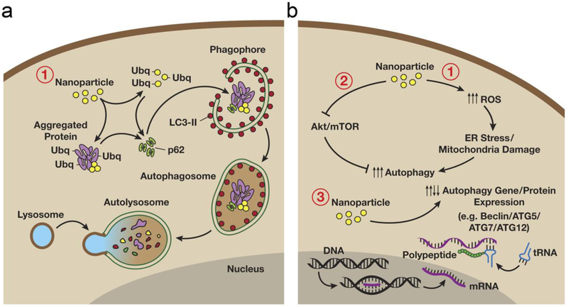
Autophagy is a conserved process that is critical for sequestering and degrading proteins, damaged or aged organelles, and for maintaining cellular homeostasis under stress conditions. Despite its dichotomous role in health and diseases, autophagy usually promotes growth and progression of advanced cancers. In this context, clinical trials using chloroquine and hydroxychloroquine as autophagy inhibitors have suggested that autophagy inhibition is a promising approach for treating advanced malignancies and/or overcoming drug resistance of small molecule therapeutics (i.e., chemotherapy and molecularly targeted therapy). Efficient delivery of autophagy inhibitors may further enhance the therapeutic effect, reduce systemic toxicity, and prevent drug resistance. As such, nanocarriers-based drug delivery systems have several distinct advantages over free autophagy inhibitors that include increased circulation of the drugs, reduced off-target systemic toxicity, increased drug delivery efficiency, and increased solubility and stability of the encapsulated drugs. With their versatile drug encapsulation and surface-functionalization capabilities, nanocarriers can be engineered to deliver autophagy inhibitors to tumor sites in a context-specific and/or tissue-specific manner. This review focuses on the role of nanomaterials utilizing autophagy inhibitors for cancer therapy, with a focus on their applications in different cancer types.

Keywords: Autophagy; cancer; drug delivery; nanomaterials; theranostics.


Conflict of interest statement

Conflicts of interest The authors declare no competing financial interest.

Figures

Figure 1.
Figure 1.
Nanoparticle-mediated autophagy. (a) Autophagy involves the sequestration and subsequent degradation of nanomaterials. After entrance into the cytoplasm, nanomaterials may be sequestered and deposited within autophagosomes, and fusion of autophagosomes with lysosomes will form autolysosomes, which will result in the breakdown and degradation of encapsulated foreign or aberrant nanomaterials. (b) Nanomaterials may also induce autophagy through upregulating oxidative stress and/or affecting the intracellular signaling pathways (e.g., Akt-mTOR signaling pathway) which may regulate the expression of genes/proteins necessary for autophagosome formation. Figure adapted with permission from.[88] Copyright © 2012 BioMed Central Ltd.
Figure 2.
Figure 2.
188Re-liposome inhibited autophagy in the tumor microenvironment and significantly suppressed tumor growth in ovarian cancer models. (a) The 188Re-Liposome treated ES2 cells (poorly differentiated ovarian clear cell carcinoma cells) were grown on glass coverslips and were transfected with LC3B-GFP vectors. Treatment with 188Re-liposome in ovarian cancer models downregulated the expression of LC3B, whereas treatment with 188Re-BMEDA did not. (b) Intraperitoneal delivery of 188Re-liposome demonstrated significant tumor killing effects in the ES2-Luc ovarian cancer model, where the tumor burden was monitored by bioluminescence imaging. Scale bar: 20 μm. Figure adapted with permission from.[99] Copyright © 2017 MDPI AG.
Figure 3.
Figure 3.
Chloroquine (CQ) enhanced the therapeutic efficacy of dendritic DTX-H40-PLA NPs in breast cancer models. (a) Schema of preparation of DTX-H40-PLA NPs. (b) Field emission scanning electron microscopy imaging of the DTX-H40-PLA NPs. (c) H40-PLA NPs induced autophagy in MCF-7 cells, as revealed by the confocal microscopic imaging, and quantification of DsRed-LC3 transfected cells by western blotting of LC3I/II protein levels. Scale bar represents 10 μm. (d) Inhibition of autophagy using CQ enhanced the tumor-suppressing effect of DTX-H40-PLA NPs in MCF-7 breast cancer models (n=5 for each treatment group). * represents P<0.05 and ** represent P<0.01. (e) Assessment of autophagy activity in tumor tissues further confirmed profound autophagy inhibition in mice treated with a combination of DTX-H40-PLA NPs and CQ. Autophagosomes were indicated by the arrows on the enlarged fluorescent images of the below images. Scale bar represents 10 μm. Figure adapted with permission from.[117] Copyright © 2014 Ivyspring international publisher.
Figure 4.
Figure 4.
Hyperbranched polymers-mediated delivery of Beclin1 siRNA and antitumor DOX inhibited drug-induced autophagy and thereby suppressed tumor growth in cervical cancer models. (a) The schematic illustration of the assembly of OEI600-PBA and HBPO and the corresponding TEM image of the core-corona nanocarrier. (b) Quantitative PCR and western blotting analyses of LC3 level in HeLa cells showed that DOX treatment led to enhancement of cellular autophagy. In comparison, delivery of Beclin1 siRNA by the nanocarriers effectively inhibited the autophagy activation induced by DOX. **p < 0.01 and ***p < 0.001 versus normal control, ##p < 0.01 and ###p < 0.001 versus DOX-treated group. (c) In tumor-bearing nude mice, DOX/siRNA/(OEI600-PBA)10 outperformed other treatment formulations and showed the most obvious antitumor effect (n=5 for each treatment group). Figure adapted with permission from.[120] Copyright © 2015 Elsevier Ltd.
Figure 5.
Figure 5.
Gold nanorods (GNRs) did not induce autophagy. (a) Incubation of normal human foreskin BJ fibroblasts with 50 μM of GNRs for 24 h did not induce autophagy. The serum-starved sample (EBSS) was used as a positive control. (b) Confocal images showed colocalization of GNRs with lysosomal marker LAMP-1 in both BJ and DU145 cells. Scale bar represents 10 μm. (c) DU145 cells were treated with 30-100 μM of GNRs for 24 h and then stained using acridine orange, a sensitive marker of lysosome intactness. While the lysosomal destabilizer (hydrogen peroxide, H2O2) induced loss of lysosomal red and increase of the cytoplasmic acridine orange in DU145 cells, GNRs did not inflict permeabilization of the lysosomes in DU145 cells. Scale bar represents 10 μm. Figure adapted with permission from.[130] Copyright © 2018 Elsevier Ltd.
Figure 6.
Figure 6.
Inhibition of autophagy enhanced the cytotoxicity of silver nanoparticles (Ag NPs) in HeLa cells. (a) TEM image of Ag NPs. (b) Size distribution of Ag NPs measured using dynamic light scattering. (c) Flow cytometry assessment of apoptosis using annexin A5-FITC (ANXA5-FITC)/propidium iodide (PI) showed extensive late-stage apoptosis of HeLa cells treated with Ag NPs and autophagy inhibitor 3-MA. (d) Reducing TFEB protein level by TFEB-specific siRNA also enhanced the cancer cell killing effect of Ag NPs. Figure adapted with permission from.[71] Copyright © 2018 John Wiley & Sons, Inc.
Figure 7.
Figure 7.
Synergistic anti-tumor and anti-metastasis effect of D/PSP@CQ/CaP NPs in 4T1 murine breast cancer models. (a) Schema illustrating mechanisms of D/PSP@CQ/CaP NPs in suppressing breast cancer in an autophagy-dependent manner. (b) Efficient autophagy inhibition by CQ/CaP shell in 4T1 breast cancer cells as revealed by fluorescence imaging. (c) Treatment of orthotopic 4T1 tumors by D/PSP@CQ/CaP inhibited the tumor growth and prolonged the median survival of mice (n=15 for each treatment group). & represents p < 0.01. Figure adapted with permission from.[139] Copyright © 2017 Elsevier Ltd.
Figure 8.
Figure 8.
Molybdenum disulfide (MoS2) nanosheets can be internalized within autophagosomes. (a) The depiction of surface-engineering and drug-loading of MoS2 nanosheets. (b) Confocal imaging of EGFP-LC3 transfected HeLa cells showed colocalization of MoS2-based nanosheets and EGFP-LC3-positive autophagosomes. (c) The colocalization of MoS2-based nanosheets and autophagosomes was also observed in MCF-7 cells. Confocal images showed colocalization of lysosomes and EGFP-LC3-positive autophagosomes in HeLa cells (d) and MCF-7 cells (e) incubated with fluorescent MoS2-based nanosheets. Figure adapted with permission from.[155] Copyright © 2018 American Chemical Society.

Similar articles

Cited by

References

    1. Holohan C, Van Schaeybroeck S, Longley DB, Johnston PG, Nat. Rev. Cancer 2013, 13, 714. - PubMed
    1. Amaravadi RK, Yu D, Lum JJ, Bui T, Christophorou MA, Evan GI, Thomas-Tikhonenko A, Thompson CB, J. Clin. Invest 2007, 117, 326. - PMC - PubMed
    1. Briceno E, Reyes S, Sotelo J, Neurosurg. Focus 2003, 14, e3. - PubMed
    1. Briceno E, Calderon A, Sotelo J, Surg. Neurol 2007, 67, 388. - PubMed
    1. Sasaki K, Tsuno NH, Sunami E, Tsurita G, Kawai K, Okaji Y, Nishikawa T, Shuno Y, Hongo K, Hiyoshi M, Kaneko M, Kitayama J, Takahashi K, Nagawa H, BMC Cancer 2010, 10, 370. - PMC - PubMed