. 2019 Jul 24;5(7):eaaw3963.
doi: 10.1126/sciadv.aaw3963. eCollection 2019 Jul.

Bioinspired mechanically active adhesive dressings to accelerate wound closure

Affiliations

Bioinspired mechanically active adhesive dressings to accelerate wound closure

S O Blacklow et al. Sci Adv. .

Abstract

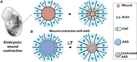
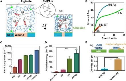
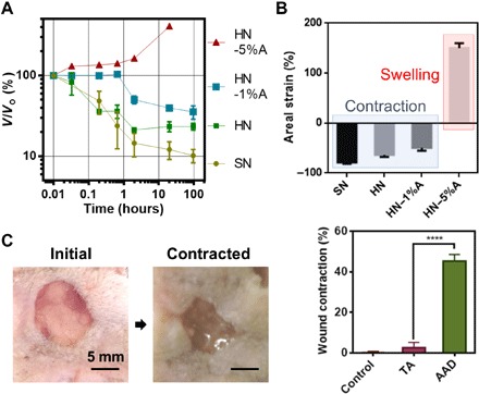
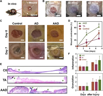
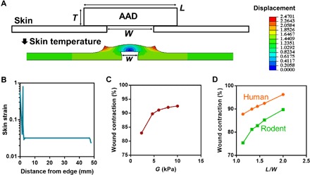
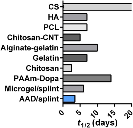
Inspired by embryonic wound closure, we present mechanically active dressings to accelerate wound healing. Conventional dressings passively aid healing by maintaining moisture at wound sites. Recent developments have focused on drug and cell delivery to drive a healing process, but these methods are often complicated by drug side effects, sophisticated fabrication, and high cost. Here, we present novel active adhesive dressings consisting of thermoresponsive tough adhesive hydrogels that combine high stretchability, toughness, tissue adhesion, and antimicrobial function. They adhere strongly to the skin and actively contract wounds, in response to exposure to the skin temperature. In vitro and in vivo studies demonstrate their efficacy in accelerating and supporting skin wound healing. Finite element models validate and refine the wound contraction process enabled by these active adhesive dressings. This mechanobiological approach opens new avenues for wound management and may find broad utility in applications ranging from regenerative medicine to soft robotics.


Figures

Fig. 1
Fig. 1. Bioinspired design of AAD for promoting wound contraction.
(A) Skin wounds of chicken embryo. An actin cable (green) is formed in the cells at the wound edges and contacts the wound. (B) Active wound contraction enabled by AAD that adheres to and contracts the wound edges at the skin temperature. Red dashed arrows indicate the contraction.
Fig. 2
Fig. 2. Mechanical and antibacterial properties of AAD.
(A) Schematics of temperature-triggered transition of AAD, which consist of PNIPAm (blue lines), alginate (black lines), and AgNPs (Ag; grey spheres). AAD forms strong adhesion (green lines) with the wounded skin. (B) Stress-strain curves of various hydrogel matrices, including the single-network PNIPAm hydrogel (SN), the hybrid-network PNIPAm-alginate hydrogel (HN), and AgNP-laden hybrid hydrogel (HN-Ag). HN were gelled at 4°C and compared to the same hydrogel form at room temperature (HN-RT). (C) Matrix toughness of various hydrogel matrices. Alg refers to alginate hydrogels and 2M refers to 2 M NIPAm monomers used to form HN. Mean ± SD, n = 3 to 5. (D) Adhesion energy measured on porcine skin. Band-Aid was included for comparison. Mean ± SD, n = 3 to 4. (E) Antimicrobial function of AAD with and without AgNPs (Ag) (bottom); a schematic of AAD and bacterial growth on agar gels (top). Mean ± SD, n = 7, ***P < 0.001.
Fig. 3
Fig. 3. Thermoresponsiveness of AAD and in vitro contraction on skin.
(A) Thermoresponsive behavior varies with the matrix composition of HN. 1%A and 5%A refer to 1% and 5% of acrylamide within the total monomer. SN represents PNIPAm-only gel used as a control. (B) Areal strains as a function of the matrix composition measured under equilibrium conditions at 37°C. Negative values of the areal strain indicate contraction, whereas positive values indicate expansion. Mean ± SD, n = 3 to 4. (C) In vitro tests of AAD-enabled wound contraction on fresh rodent skin. Comparison of the wound area between nontreatment control, nonthermoresponsive dressing TA, and AAD. Mean ± SD, n = 5, ****P < 0.0005. Photo credit: Jianyu Li and David Mooney, Harvard University.
Fig. 4
Fig. 4. In vivo wound healing with application of AAD.
(A) In vivo rodent wounds with splints. (B) Skin temperature–triggered response of AAD on a skin wound. Scale bars, 5 mm. (C) Digital images of the initial wounds and those after 7 days with no hydrogel treatment (control), with treatment with nonthermoresponsive TA, and with treatment with AAD. Scale bars, 2 mm. (D) Wound contraction as function of time and treatments. Mean ± SD, n = 5, *P < 0.05. (E) Histological sections of the wounded skin harvested on day 7 and hematoxylin and eosin–stained. The wound edges and the residues of adhesives are marked with green triangles and black arrows, respectively. (F) Histological assessment of levels of inflammation and granulation by a blinded pathologist expert (0, normal; 1, minimal; 2, mild; 3, moderate; and 4, strong). Median ± interquartile range, n = 3 to 5 per group. Photo credit: Jianyu Li and David Mooney, Harvard University.
Fig. 5
Fig. 5. Finite element simulation of AAD-enabled wound contraction.
(A) Configuration of the finite element model where AAD of thickness T, width L, and shear modulus G is attached onto a wounded skin of initial wound size W (top). After contraction, the wound size is reduced to w (bottom). The color contours map the magnitude of horizontal displacement. (B) Simulated rodent skin strain as a function of the distance from the wound edge. (C) The wound contraction, calculated by 1 − (w/W)2, is predicted to vary with the shear modulus G of AAD. (D) Modeling of contraction of human and rodent skin as a function of the length of AAD to width of the wound ratio (L/W).
Fig. 6
Fig. 6. Wound closure rate, as characterized by the time for 50% wound closure (t1/2) with various reported wound dressings.
Comparisons include AAD (data reported in Fig. 4), microgels (25), chitosan (35), dopamine-modified polyacrylamide (PAAm-Dopa) (50), gelatin (51), alginate-gelatin hydrogels (52), carbon nanotube–laden chitosan hydrogels (chitosan-CNT) (53), polycaprolactone nanofibrous matrix (PCL) (54), hyaluronan (HA), and chondroitin sulfate (CS) hydrogels (55). The half-life time (t1/2) resulting from chondroitin sulfate hydrogels was beyond 20 days. While AAD and microgels were tested in a rodent wound model with splints, most previous reports did not use splinting.

References

    1. Singer A. J., Clark R. A. F., Cutaneous wound healing. N. Engl. J. Med. 341, 738–746 (1999). - PubMed
    1. Fong E., Tirrell D. A., Collective cell migration on artificial extracellular matrix proteins containing full-length fibronectin domains. Adv. Mater. 22, 5271–5275 (2010). - PMC - PubMed
    1. Zhu S., Nih L., Carmichael S. T., Lu Y., Segura T., Enzyme-responsive delivery of multiple proteins with spatiotemporal control. Adv. Mater. 27, 3620–3625 (2015). - PMC - PubMed
    1. Dyson M., Young S., Pendle C. L., Webster D. F., Lang S. M., Comparison of the effects of moist and dry conditions on dermal repair. J. Invest. Dermatol. 91, 434–439 (1988). - PubMed
    1. Yannas I. V., Burke J. F., Orgill D. P., Skrabut E. M., Wound tissue can utilize a polymeric template to synthesize a functional extension of skin. Science 215, 174–176 (1982). - PubMed

Publication types

LinkOut - more resources