. 2019 Apr;1(4):213-226.
doi: 10.1096/fba.2018-00018. Epub 2018 Nov 29.

Inhibition of osteoclastogenesis by opsonized Porphyromonas gingivalis

Affiliations

Inhibition of osteoclastogenesis by opsonized Porphyromonas gingivalis

Subramanya N Pandruvada et al. FASEB Bioadv. 2019 Apr.

Abstract

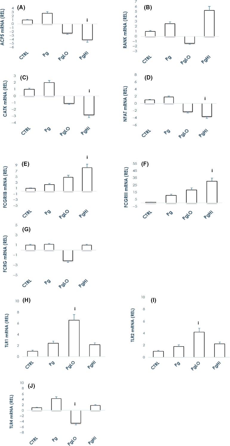
A crucial step in the pathogenesis of periodontal disease (PD) is activation of osteoclasts (OC) by numerous virulence factors produced by Porphyromonas gingivalis (Pg). To understand pathogenesis of periodontal disease and the role of specific adaptive immune responses, effects of antibodies on Pg-induced OC differentiation and function were investigated. Human peripheral blood-derived monocytes were differentiated in vitro to osteoclasts in the presence or absence of: a) Pg; b) antibodies to Pg; and c) antibody-opsonized Pg. Findings suggest significant induction of osteoclastogenesis by Pg when compared to control cultures, whereas opsonization decreased osteoclastogenesis by 45%. Immune receptor gene expression profile in the presence of opsonized Pg showed marked up-regulation of TLR1 (3-fold) and TLR2 (2-fold) along with FcγRIIB (2-fold) and FcγRIII receptors (5-fold), but not TLR4 and FcRγ receptors. Interestingly, blocking FcγRIIB, but not FcγRIII receptor, reversed the inhibitory effects of opsonized Pg suggesting a critical role played by FcγRIIB in osteoclastogenesis. Furthermore, opsonized Pg transformed OC precursors to a "macrophage phenotype" suggesting a bone protective role of the immune complexes in modulating osteoclastogenesis, probably by competing as an agonist for PRRs, and inducing selective activation of FcγRs with simultaneous suppression of FcRγ which regulates bone resorptive process. Further defining effective antibody isotypes, avidity, and antigenic specificity could improve targets for eliciting protective immunity.

Keywords: bone loss; immune response; macrophages; osteoclast; periodontitis.


Conflict of interest statement

Conflict of Interest Statement: Authors report no conflicts of interest related to this study.

Figures

Figure 1
Figure 1
Opsonized Porphyromonas gingivalis inhibits osteoclast differentiation. A, Human peripheral blood‐derived monocytes from healthy donors were cultured in the presence of macrophage colony stimulating factor (M‐CSF) and receptor activator of nuclear Factor‐kappa B ligand (RANKL) or formalin fixed or heat‐killed or live P gingivalis at various cfu/mL for 2 weeks. Osteoclasts were stained for TRAcP activity, quantified, and grouped based on number of nuclei (three or more nuclei). Multinucleated cells with three or more nuclei are counted as osteoclasts and presented as a ratio over RANKL control. Data expressed as the mean ± SE of triplicate determinations from four independent donors. i P < 0.05 vs RANKL control. B, PBMC‐derived monocytes were cultured in the presence of M‐CSF and RANKL with or without opsonized bacteria (antibodies to P gingivalis complexed with formalin fixed‐P gingivalis; Pg+abLO/Pg+abHI) and P gingivalis alone (Pg) for 12 d. Osteoclast were fixed and stained for TRAcP activity and quantified. Note increased osteoclast number with Pg alone, whereas presence of antibodies to Pg abrogates this increase significantly. Data represent the mean ± SE from three independent cultures. i < 0.05 vs RANKL control; ii < 0.05 vs Pg alone. C, PBMC‐derived monocytes were cultured in the presence of M‐CSF and RANKL (RL CTRL) or Pg (106) and/or with immune complexes (Pg+abLO/Pg+abHI) for 12 d. Osteoclast were removed by hypo‐osmotic lysis and pits were stained in Toluidine blue and were photographed. Pit area was measured using ImageJ. Histograms represent the percentage of resorbed area from three independent experiments. i < 0.05 vs Pg alone. Scale bar, 200 µm. D, Human peripheral blood‐derived monocytes from healthy donors were cultured in the presence of M‐CSF and RANKL or formalin‐fixed P gingivalis (at 106 cfu/mL); Streptococcus gordonii (1010 cfu/mL) or Pg+Sg combo for 2 weeks. Osteoclasts were stained for TRAcP activity and quantified. Histograms represent the percentage of MNC over RANKL group. Data expressed as the mean ± SE of triplicate determinations from three independent donors. i P < 0.05 vs RANKL control; ii P < 0.05 vs Pg alone. E, PBMC‐derived monocytes were cultured in the presence of M‐CSF and RANKL with or without opsonized bacteria (antibodies to P gingivalis complexed with formalin‐fixed S gordonii; Sg+abLO/Sg+abHI) and S gordonii alone (Sg) for 12 d. Osteoclast were fixed and stained for TRAcP activity and quantified. Note decreased osteoclast number with Sg treated cultures and no effect of opsonized Sg treatment. Histograms represent the percentage of MNC over RANKL group. Data expressed are the mean ± SE from three independent cultures. i P < 0.05 vs RANKL control
Figure 2
Figure 2
Opsonization of bacteria abrogates Porphyromonas gingivalis’ stimulatory effects by modulating immune receptors gene expression. PBMC‐derived monocytes were cultured in the presence of M‐CSF and RANKL for 7 d and treated with Pg or antibodies to Pg (LO/HI) alone or with opsonized bacteria (PgLO/PgHI) for 24 hours. Pre‐osteoclasts lysed in TRIzol were processed for RNA and subsequent cDNA preparations. QPCR analyses done to assay immune receptor gene expression revealed upregulation of TLR1, TLR2, FcγRIIB, and FcγRIII gene expression levels but not that of TLR4 and FcRγ when compared to Pg alone. Bars represent mean ± SE from three independent donors. i P < 0.05 vs Pg alone. Note that graphs presented are not on the same scale
Figure 3
Figure 3
Opsonization of bacteria abrogates Porphyromonas gingivalis’ stimulatory effects by modulating immune receptor expression. A, PBMC‐derived monocytes were cultured in the presence of macrophage colony stimulating factor (M‐CSF) and receptor activator of nuclear Factor‐kappa B ligand for 10 d and treated with Pg or with opsonized bacteria (PgLO/PgHI) for 48 hours. Osteoclasts lysed in RIPA buffer were processed for total protein. Western blot analyses were done to assay immune receptor expression revealed upregulation of FCγRIIB and FCγRIII, TLR1 and TLR2 expression levels but not that of TLR4 when compared to Pg alone. Expression levels were normalized to that of β‐actin. Bars represent mean ± SE from two independent experiments. Note that graphs presented above are not on the same scale. B, Human monocytes were purified from PBMC and were cultured in the presence of M‐CSF and RANKL until binucleation. Pre‐osteoclasts were incubated for 1 h with 2 µg/mL blocking antibodies of FcγRII/FcγRIII/IgG isotype. Subsequently, treated with P gingivalis alone (Pg) or opsonized bacteria (antibodies to P gingivalis complexed with formalin fixed‐P gingivalis: PgLO/PgHI) until multinucleation. Osteoclasts were fixed and stained for TRAcP activity and quantified. Bars represent mean ± SE from two independent cultures. Note reversal of inhibitory effects of opsonized Pg following inhibition of FcγRII receptor whereas significant decrease in osteoclast number following application of opsonized bacteria after blocking FcγRIII. i P < 0.05 vs Pg alone. Bars represent mean ± SE from two independent donors
Figure 4
Figure 4
Opsonization of bacteria abrogates Porphyromonas gingivalis’ stimulatory effects by driving preosteoclasts to “M2 like” phenotype. A, PBMC‐derived monocytes were cultured in the presence of macrophage colony stimulating factor (M‐CSF) and receptor activator of nuclear Factor‐kappa B ligand for 3 d and treated with Pg or with opsonized bacteria (PgLO/PgHI) for 48 h. Cell surface antigens on preosteoclasts were processed for flow cytometry. Briefly, preosteoclasts were rinsed in PBS and detached using PBS‐EDTA. Detached cells were washed in PBS and blocked in PBS‐BSA. Cell surface antibody analysis done to assay macrophage marker expression revealed upregulation of CD68 and CD168 with marked downregulation of CD80 in opsonized Pg treated group. Expression levels were normalized to that of IgG Isotype control. Values represent percentage of positive cells (mean ± SE) from two independent donors. i P < 0.05 vs Pg alone. The data shown are representative flow cytometry analysis from one of two independent experiments. B, PBMC‐derived monocytes were cultured in the presence of M‐CSF and RANKL for 3 d and treated with Pg either free or opsonized (PgLO/PgHI) for 24 h. Pre‐osteoclasts lysed in TRIzol were processed for RNA and subsequent cDNA preparations. QPCR analyses to assay macrophage signature gene expression revealed upregulation of CCL17, CCL22, KLF4, and MRC1 (M2 genes) but not that of CXCL10 and CXCL11 (M1 genes) when compared to Pg alone. Bars represent mean ± SE from two independent donors. i P < 0.05 vs Pg alone. Note that graphs presented are not on the same scale
Figure 5
Figure 5
Hypothetical mechanisms of adaptive immune response modulating osteoclastic bone resorption in periodontal diseases. Porphyromonas gingivalis both directly, and indirectly through B and T cell stimulation promote osteoclast differentiation and contribute to periodontal bone loss. Interestingly, Pg primes antigen specific B cells for antibody production and the IgG antibody response to Pg induced by infection/immunization which are delivered to the gingiva by the vasculature, may result in opsonization of Pg. Our findings suggest that these opsonized Pg block OC differentiation by interacting with TLR and Fc receptors. FcγR being an inhibitory receptor under physiological conditions, is downregulated as osteoclastogenesis proceeds, resulting in an increased cell surface expression of OSCAR due to increased availability of FcRγ. FcRγ, the signaling adaptor molecule for OSCAR, is also shared by IgG‐Fc receptors (FcγRIA and FcγRIII). As influenced by the interaction of the osteoclastic IgG‐Fc receptors with immune complexes (IC), co‐stimulatory signaling (dotted lines) results in inhibition of osteoclastogenesis. Our findings support the idea that opsonized‐Pg induce upregulation of inhibitory FcγRs and enhanced TLR signaling, thereby attenuating RANK and co‐stimulatory FcRγ‐induced osteoclastogenesis (dotted lines in red)

Similar articles

Cited by

References

    1. Kassebaum NJ, Bernabe E, Dahiya M, Bhandari B, Murray CJ, Marcenes W. Global burden of severe periodontitis in 1990–2010: a systematic review and meta‐regression. J Dent Res. 2014;93:1045‐1053. - PMC - PubMed
    1. Cochran DL. Inflammation and bone loss in periodontal disease. J Periodontol. 2008;79:1569‐1576. - PubMed
    1. Hienz SA, Paliwal S, Ivanovski S. Mechanisms of bone resorption in periodontitis. J Immunol Res. 2015;2015:615486. - PMC - PubMed
    1. Holt SC, Ebersole JL. Porphyromonas gingivalis, Treponema denticola, and Tannerella forsythia: the “red complex”, a prototype polybacterial pathogenic consortium in periodontitis. Periodontol 2000. 2005;38:72‐122. - PubMed
    1. Ebersole JL. Humoral immune responses in gingival crevice fluid: local and systemic implications. Periodontol 2000. 2003;31:135‐166. - PubMed

LinkOut - more resources