Review
. 2019 Jul;290(1):60-84.
doi: 10.1111/imr.12773.

CAR T cells for brain tumors: Lessons learned and road ahead

Affiliations
Review

CAR T cells for brain tumors: Lessons learned and road ahead

David Akhavan et al. Immunol Rev. 2019 Jul.

Abstract

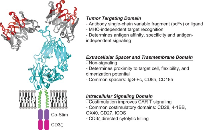
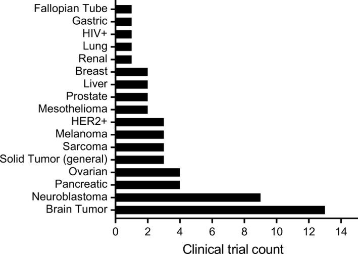
Malignant brain tumors, including glioblastoma, represent some of the most difficult to treat of solid tumors. Nevertheless, recent progress in immunotherapy, across a broad range of tumor types, provides hope that immunological approaches will have the potential to improve outcomes for patients with brain tumors. Chimeric antigen receptors (CAR) T cells, a promising immunotherapeutic modality, utilizes the tumor targeting specificity of any antibody or receptor ligand to redirect the cytolytic potency of T cells. The remarkable clinical response rates of CD19-targeted CAR T cells and early clinical experiences in glioblastoma demonstrating safety and evidence for disease modifying activity support the potential of further advancements ultimately providing clinical benefit for patients. The brain, however, is an immune specialized organ presenting unique and specific challenges to immune-based therapies. Remaining barriers to be overcome for achieving effective CAR T cell therapy in the central nervous system (CNS) include tumor antigenic heterogeneity, an immune-suppressive microenvironment, unique properties of the CNS that limit T cell entry, and risks of immune-based toxicities in this highly sensitive organ. This review will summarize preclinical and clinical data for CAR T cell immunotherapy in glioblastoma and other malignant brain tumors, including present obstacles to advancement.

Keywords: T cells; brain tumors; chimeric antigen receptors; glioblastoma.


Conflict of interest statement

Patents associated with CAR design, T cell manufacturing, and delivery have been licensed by Mustang Bio., Inc, for which CEB receives licensing and consulting payments.

Figures

Figure 1
Figure 1
Chimeric antigen receptor (CAR) design. CARs are modular synthetic immunoreceptors that consist of a tumor targeting domain fused to an intracellular T cell signaling domain via the extracellular spacer and transmembrane (TM) domains. The tumor targeting domain has been designed and tested against multiple brain tumor antigens including IL13Rα2, HER2, and EGFRvIII (Table 2). The TM domain and extracellular spacer influence effector‐target cell interaction by providing flexibility, allowing dimerization to occur, and influencing stability. The cytoplasmic intracellular signaling domain is composed of a CD3ζ activation domain, and is most often paired with cognate T‐cell co‐stimulatory signaling domains (CD28, 4‐1BB, OX40, CD27, and ICOS), which improves CAR T‐cell proliferation, survival, and recursive killing
Figure 2
Figure 2
Chimeric antigen receptor (CAR) T cell trials for brain tumors vs other solid tumors. Clinical trial count in the United States evaluating CAR T cell therapy for solid tumors as of April 2019. As graphically represented, the largest number of CAR T cell trials for solid tumors is for brain tumors, which include trials for glioblastoma and other malignant gliomas (n = 12), as well as brain metastases (n = 1). Listed trials include those that are pending activation, enrolling patients, and completed
Figure 3
Figure 3
Chimeric antigen receptor (CAR) T cell distribution following locoregional delivery. Primary glioblastoma cells (1 × 105 PBT030‐2) were implanted intracranially (IC) in the left hemisphere (2.0 mm lateral, 0.5 mm anterior to the bregma, 2.15‐3.0 mm depth from dura) of NSG mice, and 8 d later 89Zr‐oxine labeled IL13Rα2‐CAR T cells (2 × 106) were administered either IC in the right hemisphere, or into the right ventricle (ICV; 0.9 mm lateral, 0.3 mm caudal to the bregma, 2.5 mm depth from dura). Representative PET images are depicted at 15 min, 6 h, 1 d or 5 d after ICT (top) or ICV (bottom) delivery of 89Zr‐oxine labeled IL13Rα2‐CAR T cells are depicted. Color scale indicates percentages of injected radioactive dose (ID) per gram weight of voxel that were calculated using Vivo‐Quant (Invicro) analysis of the PET images205

References

    1. Siegel RL, Miller KD, Jemal A. Cancer statistics, 2018. CA Cancer J Clin. 2018;68(1):7‐30. - PubMed
    1. Stupp R, Taillibert S, Kanner A, et al. Effect of tumor‐treating fields plus maintenance temozolomide vs maintenance temozolomide alone on survival in patients with glioblastoma: a randomized clinical trial. JAMA. 2017;318(23):2306‐2316. - PMC - PubMed
    1. Alexander BM, Cloughesy TF. Adult glioblastoma. J Clin Oncol. 2017;35(21):2402‐2409. - PubMed
    1. Pollack IF, Agnihotri S, Broniscer A. Childhood brain tumors: current management, biological insights, and future directions. J Neurosurg Pediatr. 2019;23(3):261‐273. - PMC - PubMed
    1. Sperduto PW, Kased N, Roberge D, et al. Summary report on the graded prognostic assessment: an accurate and facile diagnosis‐specific tool to estimate survival for patients with brain metastases. J Clin Oncol. 2012;30(4):419‐425. - PMC - PubMed

Publication types

Substances