Review
. 2019 Jul;290(1):127-147.
doi: 10.1111/imr.12772.

T cell receptor-based cancer immunotherapy: Emerging efficacy and pathways of resistance

Affiliations
Review

T cell receptor-based cancer immunotherapy: Emerging efficacy and pathways of resistance

Smita S Chandran et al. Immunol Rev. 2019 Jul.

Abstract

Adoptive cell transfer (ACT) using chimeric antigen receptor (CAR)-modified T cells can induce durable remissions in patients with refractory B-lymphoid cancers. By contrast, results applying CAR-modified T cells to solid malignancies have been comparatively modest. Alternative strategies to redirect T cell specificity and cytolytic function are therefore necessary if ACT is to serve a greater role in human cancer treatments. T cell receptors (TCRs) are antigen recognition structures physiologically expressed by all T cells that have complementary, and in some cases superior, properties to CARs. Unlike CARs, TCRs confer recognition to epitopes derived from proteins residing within any subcellular compartment, including the membrane, cytoplasm and nucleus. This enables TCRs to detect a broad universe of targets, such as neoantigens, cancer germline antigens, and viral oncoproteins. Moreover, because TCRs have evolved to efficiently detect and amplify antigenic signals, these receptors respond to epitope densities many fold smaller than required for CAR-signaling. Herein, we summarize recent clinical data demonstrating that TCR-based immunotherapies can mediate regression of solid malignancies, including immune-checkpoint inhibitor refractory cancers. These trials simultaneously highlight emerging mechanisms of TCR resistance. We conclude by discussing how TCR-based immunotherapies can achieve broader dissemination through innovations in cell manufacturing and non-viral genome integration techniques.

Keywords: CRISPR/Cas9; ImmTAC; TCR mimic; adoptive immunotherapy; genetic engineering.


Conflict of interest statement

Advisory/consulting: Aleta BioTherapeutics, Bellicum Pharmaceuticals, BMS, Cell Design Labs, G1 Therapeutics, Klus Pharma, Obsidian Therapeutics, Rxi Therapeutics (CAK); Honoraria: Kite/Gilead (CAK); Clinical research support: Kite/Gilead (CAK). CAK and SSC hold provisional patents related to T cell therapy and TCR engineering.

Figures

Figure 1
Figure 1
T cell receptors (TCRs) recognize a larger universe of protein‐derived antigens compared with chimeric antigen receptors (CARs). Membrane‐associated proteins, which collectively represent ~27% of the human proteome, can be targeted by both CAR and TCR‐based immunotherapies. TCRs, by contrast, can also target intracellular targets, including cytoplasmic and intra‐nuclear proteins. TCR epitopes are derived from a diverse variety of antigen classes, including tissue‐differentiation antigens, cancer germline antigens, viral oncoproteins, and mutated antigens (cancer neoantigens)
Figure 2
Figure 2
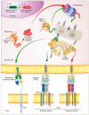
Processing, presentation, and detection of cancer‐associated antigens by chimeric antigen receptors (CARs) and T cell receptors (TCRs). Antigen processing and presentation is a continuous process in most nucleated cells, including cancer cells. This process starts with the transcription of genes encoding proteins that may be destined either for the cell surface membrane (eg, mesothelin) or the cytosol (eg, NY‐ESO‐1). Ribosomes translate transcribed RNA into proteins in the cytoplasm which are then shuttled directly into the endoplasmic reticulum (ER) for proper folding or into the proteasome for degradation. The proteasome generates linear peptide fragments that are transported into the ER via a transporter associated with antigen presentation and loaded on to a major histocompatibility complex (MHC) molecule. Newly folded surface proteins and loaded peptide/MHC (pMHC) complexes then move into the Golgi complex where they are exported to the cell surface. CARs exclusively recognize cell surface proteins; distinct TCRs can recognize pMHC complexes derived from either the cell surface or intracellular proteins. MSLN = gene encoding Mesothelin, CTAG1A = gene encoding NY‐ESO‐1; VL = variable light chain; VH = variable heavy chain; ‐SS‐ = disulfide bond
Figure 3
Figure 3
Viral transfer of an exogenous cancer‐specific T cell receptor (TCR) and TCR modifications to enhance both safety and functionality. Double stranded (ds) DNA encoding an exogenous TCR are randomly integrated into the genome of a donor T cell by viral vectors, including γ‐retroviral (shown) and lentiviral vectors. The endogenous α/β TCR, encoded by the TCR locus, remains intact and continues to be expressed. As a result, multiple TCR heterodimers may be expressed on an individual T cell's surface, including a mixture of properly paired endogenous (α/β) and exogenous (α’/β’) TCRs as well as mispaired (α/β’ or α’/β) TCRs incorporating chains from both receptors. Mispaired TCRs can reduce the function of properly paired TCRs through competition for signaling molecules, such as CD3ζ, and may also generate new specificities capable of inducing graft versus host disease. The structure of the exogenous TCR may be modified in a variety of ways to either enhance safety and/or augment function. These include: substitution of all or selected murine residues in place of the human sequence in the TCR constant regions (murinization); addition of an extra cysteine residue to promote a second disulfide bond (cysteine‐modification); modification of the hydrophobicity of the TCRα TM region (transmembrane‐modification); inversion of the human sequences of the TCR α and β constant domains (domain‐swapping); mutagenesis of the complementarity‐determining regions loops (affinity‐enhancement); consolidation of a normal TCR heterodimer into a single‐chain format by covalently linking the variable domains of the TCR chains (single‐chain). V = variable region; C = constant region; MC = murinized constant region; RT = reverse transcriptase; ‐SS‐ = disulfide bond
Figure 4
Figure 4
Targeted T cell receptor (TCR) delivery and TCR‐like structures. CRISPR/Cas9‐mediated TCR delivery can direct the targeted genomic replacement of an exogenous TCR into the endogenous TRAC locus. Disruption of the endogenous TCR eliminates expression of endogenous and mispaired TCRs. The exogenous TCR is homogenously and stably expressed under the endogenous TRAC promoter on the cell surface. TCR‐like structures, including bispecific soluble TCRs (immune‐mobilizing monoclonal TCRs against cancer; ImmTAC), bispecific antibody‐like structures (Bispecific T‐cell Engagers; BiTE), and antibodies specific for peptide/MHC complexes (TCR mimics) provide alternative approaches to re‐direct T cell specificities to tumor antigens without the need for genomic integration. V = variable region; C = constant region; VL = variable light chain; VH = variable heavy chain; CL = constant light chain; CH = constant heavy chain ‐SS‐ = disulfide bond
Figure 5
Figure 5
Mechanisms of therapeutic resistance to T cell receptor (TCR)‐based cancer immunotherapies. The mechanistic basis for TCR resistance can be subdivided into the following four categories: primary (1°) versus late/acquired and T cell‐intrinsic versus extrinsic. Strategies to successfully overcome resistance to TCR‐based immunotherapies are possible and are focused on the specific resistance category into which a patient falls

References

    1. Ribas A, Wolchok JD. Cancer immunotherapy using checkpoint blockade. Science. 2018;359(6382):1350‐1355. - PMC - PubMed
    1. Samstein RM, Lee C‐H, Shoushtari AN, et al. Tumor mutational load predicts survival after immunotherapy across multiple cancer types. Nat Genet. 2019;51(2):202‐206. - PMC - PubMed
    1. Siegel RL, Miller KD, Jemal A. Cancer statistics, 2019. CA Cancer J Clin. 2019;69(1):7‐34. - PubMed
    1. van Rooij N, van Buuren MM, Philips D, et al. Tumor exome analysis reveals neoantigen‐specific T‐cell reactivity in an ipilimumab‐responsive melanoma. J Clin Oncol. 2013;31(32):e439‐442. - PMC - PubMed
    1. Robbins PF, Lu Y‐C, El‐Gamil M, et al. Mining exomic sequencing data to identify mutated antigens recognized by adoptively transferred tumor‐reactive T cells. Nat Med. 2013;19(6):747‐752. - PMC - PubMed

Publication types

MeSH terms

Substances