. 2019 Nov 4;152(6):706-717.
doi: 10.1093/ajcp/aqz089.

Metastatic Basal Cell Carcinoma

Affiliations

Metastatic Basal Cell Carcinoma

Alvaro C Laga et al. Am J Clin Pathol. .

Abstract

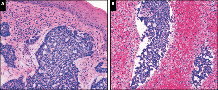
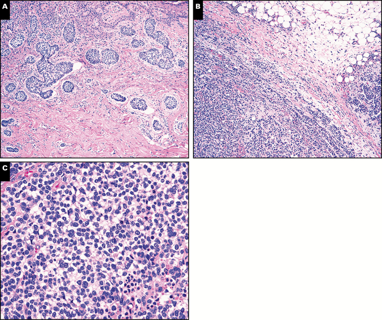
Objectives: Diagnosis of metastatic basal cell carcinoma (BCC) remains challenging, in part due to its rarity. With the advent of molecularly targeted therapies, recognition of this entity is more important than ever.

Methods: We identified 11 cases of metastatic BCC over a 13-year period. We analyzed these tumors in conjunction with their respective primary tumors by histomorphologic, immunohistochemical, and molecular genetic analyses.

Results: We identified three morphologic patterns of metastasis in BCC. The most common (seven cases) was characterized by completely typical features of BCC. Two cases showed marked squamous differentiation within BCC. The final two cases showed exclusively features of a poorly differentiated carcinoma. One of these was definitively classified by molecular analysis, as both the primary and metastatic tumors harbored the same inactivating PTCH1 mutation.

Conclusions: This study illustrates multiple distinct morphologic patterns in metastatic BCC and highlights the utility of ancillary molecular testing for accurate diagnosis.

Keywords: Basal cell carcinoma; Bone; Lung; Lymph node; Metastasis.


Figures

Image 1
Image 1
A, Case 2. Typical features of basal cell carcinoma (BCC) in the primary skin tumor (H&E, x200). B, Case 2. Typical features of BCC in the lymph node metastasis. Prominent central necrosis is noted in the metastasis, a feature present in other cases of metastatic BCC in this series (H&E, x200).
Image 2
Image 2
A, Case 5. Typical features of basal cell carcinoma (BCC) in the primary skin tumor (H&E, x400). B, Case 5. Typical features of BCC are also present in the cell block preparation from a fine-needle aspiration of the lung metastasis (H&E, x400).
Image 3
Image 3
A, Case 6. Typical features of basal cell carcinoma (BCC) are evident in the primary skin tumor (H&E, x200). B, Case 6. Lymph node metastasis with typical features of BCC (H&E, x400). C, Case 6. Typical features of BCC in bone metastasis (H&E, x400).
Image 4
Image 4
A, Case 8. Soft tissue metastasis showing prominent squamous differentiation with keratinization (H&E, x200). B, Case 8. Other areas within the same metastatic lesion show typical features of basal cell carcinoma (H&E, x200).
Image 5
Image 5
A, Case 10. Primary basal cell carcinoma (BCC) with an area of lesser differentiation (single asterisk) and another area of a poorly differentiated tumor (double asterisk) (H&E, x200). B, Case 10. Higher magnification of a poorly differentiated area (double asterisk above), not recognizable as BCC (H&E, x600). C, Case 10. Lymph node metastasis showing diffuse nodal involvement (H&E, x100). D, Case 10. Higher magnification of lymph node metastasis showing a poorly differentiated neoplasm (H&E, x600). E, Case 10. The lymph node metastasis diffusely expresses p63 by immunohistochemistry (x200).
Image 6
Image 6
A, Case 11. Primary basal cell carcinoma (BCC) showing a micronodular pattern (H&E, x200). B, Case 11. In contrast, the metastasis (right axillary lymph nodes) shows a poorly differentiated carcinoma (H&E, x200). C, Case 11. The metastasis shows sheets of large, discohesive, and hyperchromatic cells with a “clear cell” appearance, which is not recognizable as metastatic BCC (H&E, x600).
Figure 1
Figure 1
Case 11. An identical PTCH1 nonsense mutation (c.1726C > T;p.Q576*) is present in both the primary (A, B) and the metastasis (C, D), leading to biallelic genomic PTCH1 inactivation in the metastasis (A, C). An increased number of C > T transitions, enriched at dipyrimidine (CC, CT, TC) sites, characteristic of UV-induced DNA damage was identified in both tumors (B, D).

References

    1. Wu S, Han J, Li WQ, et al. . Basal-cell carcinoma incidence and associated risk factors in U.S. women and men. Am J Epidemiol. 2013;178:890-897. - PMC - PubMed
    1. Cotran RS. Metastasizing basal cell carcinomas. Cancer. 1961;14:1036-1040. - PubMed
    1. Domarus V, Stevens PJ. Metastatic basal cell carcinoma. J Am Acad Dermatol 1984;10:1043-1060. - PubMed
    1. Lo JS, Snow SN, Reizner GT, et al. . Metastatic basal cell carcinoma: report of twelve cases with a review of the literature. J Am Acad Dermatol. 1991;24:715-719. - PubMed
    1. Snow SN, Sahl W, Lo JS, et al. . Metastatic basal cell carcinoma: report of five cases. Cancer. 1994;73:328-335. - PubMed

Publication types